3PNT | pdb_00003pnt

Crystal Structure of the Streptococcus pyogenes NAD+ glycohydrolase SPN in complex with IFS, the Immunity Factor for SPN


Experimental Data Snapshot

  • Method: X-RAY DIFFRACTION
  • Resolution: 2.80 Å
  • R-Value Free: 
    0.278 (Depositor), 0.280 (DCC) 
  • R-Value Work: 
    0.208 (Depositor), 0.216 (DCC) 
  • R-Value Observed: 
    0.212 (Depositor) 

wwPDB Validation 3D Report Full Report

Validation slider image for 3PNT

This is version 1.2 of the entry. See complete history

Literature

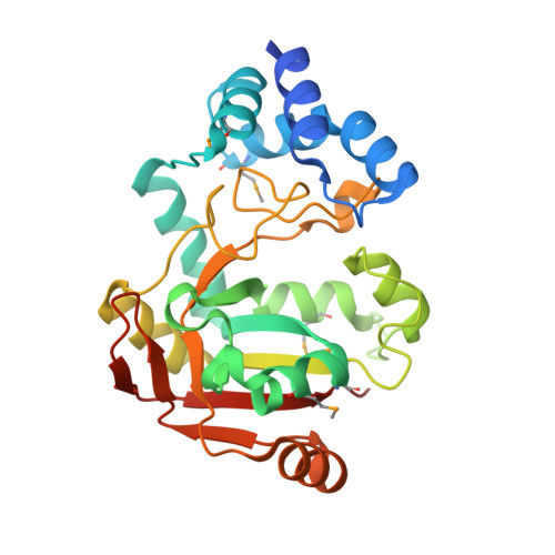
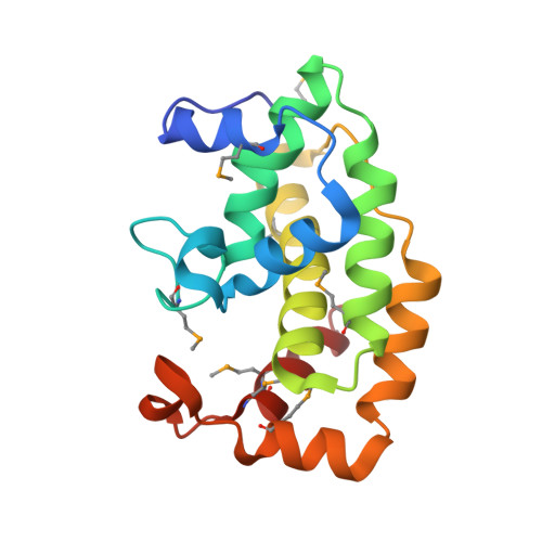
Structural Basis of Streptococcus pyogenes Immunity to Its NAD(+) Glycohydrolase Toxin.

Smith, C.L.Ghosh, J.Elam, J.S.Pinkner, J.S.Hultgren, S.J.Caparon, M.G.Ellenberger, T.

(2011) Structure 19: 192-202

  • DOI: https://doi.org/10.1016/j.str.2010.12.013
  • Primary Citation Related Structures: 
    3PNT, 3QB2

  • PubMed Abstract: 

    The virulence of Gram-positive bacteria is enhanced by toxins like the Streptococcus pyogenes β-NAD(+) glycohydrolase known as SPN. SPN-producing strains of S. pyogenes additionally express the protein immunity factor for SPN (IFS), which forms an inhibitory complex with SPN. We have determined crystal structures of the SPN-IFS complex and IFS alone, revealing that SPN is structurally related to ADP-ribosyl transferases but lacks the canonical binding site for protein substrates. SPN is instead a highly efficient glycohydrolase with the potential to deplete cellular levels of β-NAD(+). The protective effect of IFS involves an extensive interaction with the SPN active site that blocks access to β-NAD(+). The conformation of IFS changes upon binding to SPN, with repacking of an extended C-terminal α helix into a compact shape. IFS is an attractive target for the development of novel bacteriocidal compounds functioning by blocking the bacterium's self-immunity to the SPN toxin.


  • Organizational Affiliation
    • Department of Biochemistry and Molecular Biophysics, Washington University School of Medicine, Saint Louis, MO 63110-1093, USA.

Macromolecule Content 

  • Total Structure Weight: 100.76 kDa 
  • Atom Count: 6,793 
  • Modeled Residue Count: 821 
  • Deposited Residue Count: 858 
  • Unique protein chains: 2

Macromolecules

Find similar proteins by:|  3D Structure
Entity ID: 1
MoleculeChains  Sequence LengthOrganismDetailsImage
NAD+-glycohydrolase
A, C
268Streptococcus pyogenesMutation(s): 0 
Gene Names: spn
EC: 3.2.2.5
UniProt
Find proteins for D7S065 (Streptococcus pyogenes)
Explore D7S065 
Go to UniProtKB:  D7S065
Entity Groups
Sequence Clusters30% Identity50% Identity70% Identity90% Identity95% Identity100% Identity
UniProt GroupD7S065
Sequence Annotations
Expand
Reference Sequence
Find similar proteins by:|  3D Structure
Entity ID: 2
MoleculeChains  Sequence LengthOrganismDetailsImage
Immunity factor for SPN
B, D
161Streptococcus pyogenesMutation(s): 0 
Gene Names: ifs
UniProt
Find proteins for Q2VJ58 (Streptococcus pyogenes)
Explore Q2VJ58 
Go to UniProtKB:  Q2VJ58
Entity Groups
Sequence Clusters30% Identity50% Identity70% Identity90% Identity95% Identity100% Identity
UniProt GroupQ2VJ58
Sequence Annotations
Expand
Reference Sequence

Small Molecules

Modified Residues  1 Unique
IDChains TypeFormula2D DiagramParent
MSE
Query on MSE
A, C
L-PEPTIDE LINKINGC5 H11 N O2 SeMET

Experimental Data & Validation

Experimental Data

  • Method: X-RAY DIFFRACTION
  • Resolution: 2.80 Å
  • R-Value Free:  0.278 (Depositor), 0.280 (DCC) 
  • R-Value Work:  0.208 (Depositor), 0.216 (DCC) 
  • R-Value Observed: 0.212 (Depositor) 
Space Group: C 1 2 1
Unit Cell:
Length ( Å )Angle ( ˚ )
a = 198.817α = 90
b = 57.892β = 107.2
c = 89.699γ = 90
Software Package:
Software NamePurpose
REFMACrefinement
PDB_EXTRACTdata extraction

Structure Validation

View Full Validation Report


Entry History 

Deposition Data

Revision History  (Full details and data files)

  • Version 1.0: 2011-03-02
    Type: Initial release
  • Version 1.1: 2011-07-13
    Changes: Version format compliance
  • Version 1.2: 2024-11-27
    Changes: Data collection, Database references, Derived calculations, Refinement description, Structure summary