3PMQ | pdb_00003pmq

Crystal structure of the outer membrane decaheme cytochrome MtrF


Experimental Data Snapshot

  • Method: X-RAY DIFFRACTION
  • Resolution: 3.20 Å
  • R-Value Free: 
    0.321 (Depositor), 0.324 (DCC) 
  • R-Value Work: 
    0.305 (Depositor), 0.304 (DCC) 
  • R-Value Observed: 
    0.306 (Depositor) 

wwPDB Validation 3D Report Full Report

Validation slider image for 3PMQ

Ligand Structure Quality Assessment 


This is version 1.3 of the entry. See complete history

Literature

Structure of a bacterial cell surface decaheme electron conduit.

Clarke, T.A.Edwards, M.J.Gates, A.J.Hall, A.White, G.F.Bradley, J.Reardon, C.L.Shi, L.Beliaev, A.S.Marshall, M.J.Wang, Z.Watmough, N.J.Fredrickson, J.K.Zachara, J.M.Butt, J.N.Richardson, D.J.

(2011) Proc Natl Acad Sci U S A 108: 9384-9389

  • DOI: https://doi.org/10.1073/pnas.1017200108
  • Primary Citation Related Structures: 
    3PMQ

  • PubMed Abstract: 

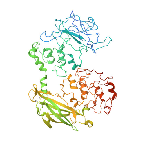
    Some bacterial species are able to utilize extracellular mineral forms of iron and manganese as respiratory electron acceptors. In Shewanella oneidensis this involves decaheme cytochromes that are located on the bacterial cell surface at the termini of trans-outer-membrane electron transfer conduits. The cell surface cytochromes can potentially play multiple roles in mediating electron transfer directly to insoluble electron sinks, catalyzing electron exchange with flavin electron shuttles or participating in extracellular intercytochrome electron exchange along "nanowire" appendages. We present a 3.2-Å crystal structure of one of these decaheme cytochromes, MtrF, that allows the spatial organization of the 10 hemes to be visualized for the first time. The hemes are organized across four domains in a unique crossed conformation, in which a staggered 65-Å octaheme chain transects the length of the protein and is bisected by a planar 45-Å tetraheme chain that connects two extended Greek key split β-barrel domains. The structure provides molecular insight into how reduction of insoluble substrate (e.g., minerals), soluble substrates (e.g., flavins), and cytochrome redox partners might be possible in tandem at different termini of a trifurcated electron transport chain on the cell surface.


  • Organizational Affiliation
    • Centre for Molecular and Structural Biochemistry, School of Biological Sciences and School of Chemistry, University of East Anglia, Norwich NR4 7TJ, United Kingdom.

Macromolecule Content 

  • Total Structure Weight: 77.08 kDa 
  • Atom Count: 4,826 
  • Modeled Residue Count: 593 
  • Deposited Residue Count: 669 
  • Unique protein chains: 1

Macromolecules

Find similar proteins by:|  3D Structure
Entity ID: 1
MoleculeChains  Sequence LengthOrganismDetailsImage
Decaheme cytochrome c MtrF669Shewanella oneidensis MR-1Mutation(s): 0 
Membrane Entity: Yes 
UniProt
Find proteins for Q8EG32 (Shewanella oneidensis (strain ATCC 700550 / JCM 31522 / CIP 106686 / LMG 19005 / NCIMB 14063 / MR-1))
Explore Q8EG32 
Go to UniProtKB:  Q8EG32
Entity Groups
Sequence Clusters30% Identity50% Identity70% Identity90% Identity95% Identity100% Identity
UniProt GroupQ8EG32
Sequence Annotations
Expand
Reference Sequence

Small Molecules

Ligands 2 Unique
IDChains Name / Formula / InChI Key2D Diagram3D Interactions
HEC

Query on HEC


Download:Ideal Coordinates CCD File
B [auth A]
C [auth A]
D [auth A]
E [auth A]
F [auth A]
B [auth A],
C [auth A],
D [auth A],
E [auth A],
F [auth A],
G [auth A],
H [auth A],
I [auth A],
J [auth A],
K [auth A]
HEME C
C34 H34 Fe N4 O4
HXQIYSLZKNYNMH-LJNAALQVSA-N
CA

Query on CA


Download:Ideal Coordinates CCD File
L [auth A]CALCIUM ION
Ca
BHPQYMZQTOCNFJ-UHFFFAOYSA-N

Experimental Data & Validation

Experimental Data

  • Method: X-RAY DIFFRACTION
  • Resolution: 3.20 Å
  • R-Value Free:  0.321 (Depositor), 0.324 (DCC) 
  • R-Value Work:  0.305 (Depositor), 0.304 (DCC) 
  • R-Value Observed: 0.306 (Depositor) 
Space Group: I 2 3
Unit Cell:
Length ( Å )Angle ( ˚ )
a = 256.64α = 90
b = 256.64β = 90
c = 256.64γ = 90
Software Package:
Software NamePurpose
ADSCdata collection
SHELXSphasing
PHENIXrefinement
MOSFLMdata reduction
SCALAdata scaling

Structure Validation

View Full Validation Report


Ligand Structure Quality Assessment 


Entry History 

Deposition Data

Revision History  (Full details and data files)

  • Version 1.0: 2011-05-25
    Type: Initial release
  • Version 1.1: 2011-07-13
    Changes: Version format compliance
  • Version 1.2: 2023-05-31
    Changes: Database references, Derived calculations
  • Version 1.3: 2024-10-30
    Changes: Data collection, Structure summary