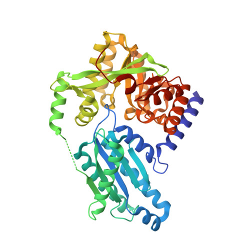
Structural Basis for c-di-GMP-Mediated Inside-Out Signaling Controlling Periplasmic Proteolysis.
Navarro, M.V., Newell, P.D., Krasteva, P.V., Chatterjee, D., Madden, D.R., O'Toole, G.A., Sondermann, H.(2011) PLoS Biol 9: e1000588-e1000588
- PubMed: 21304926
- DOI: https://doi.org/10.1371/journal.pbio.1000588
- Primary Citation of Related Structures:
3PJT, 3PJU, 3PJV, 3PJW, 3PJX - PubMed Abstract:
The bacterial second messenger bis-(3'-5') cyclic dimeric guanosine monophosphate (c-di-GMP) has emerged as a central regulator for biofilm formation. Increased cellular c-di-GMP levels lead to stable cell attachment, which in Pseudomonas fluorescens requires the transmembrane receptor LapD. LapD exhibits a conserved and widely used modular architecture containing a HAMP domain and degenerate diguanylate cyclase and phosphodiesterase domains. c-di-GMP binding to the LapD degenerate phosphodiesterase domain is communicated via the HAMP relay to the periplasmic domain, triggering sequestration of the protease LapG, thus preventing cleavage of the surface adhesin LapA. Here, we elucidate the molecular mechanism of autoinhibition and activation of LapD based on structure-function analyses and crystal structures of the entire periplasmic domain and the intracellular signaling unit in two different states. In the absence of c-di-GMP, the intracellular module assumes an inactive conformation. Binding of c-di-GMP to the phosphodiesterase domain disrupts the inactive state, permitting the formation of a trans-subunit dimer interface between adjacent phosphodiesterase domains via interactions conserved in c-di-GMP-degrading enzymes. Efficient mechanical coupling of the conformational changes across the membrane is realized through an extensively domain-swapped, unique periplasmic fold. Our structural and functional analyses identified a conserved system for the regulation of periplasmic proteases in a wide variety of bacteria, including many free-living and pathogenic species.
- Department of Molecular Medicine, College of Veterinary Medicine, Cornell University, Ithaca, New York, United States of America.
Organizational Affiliation:


