3PJX | pdb_00003pjx

Structure of Pseudomonas fluorescence LapD GGDEF-EAL dual domain, P32


Experimental Data Snapshot

  • Method: X-RAY DIFFRACTION
  • Resolution: 2.00 Å
  • R-Value Free: 
    0.222 (Depositor), 0.218 (DCC) 
  • R-Value Work: 
    0.181 (Depositor), 0.177 (DCC) 
  • R-Value Observed: 
    0.183 (Depositor) 

wwPDB Validation 3D Report Full Report

Validation slider image for 3PJX

This is version 1.2 of the entry. See complete history

Literature

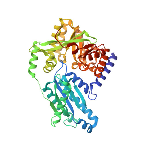
Structural Basis for c-di-GMP-Mediated Inside-Out Signaling Controlling Periplasmic Proteolysis.

Navarro, M.V.Newell, P.D.Krasteva, P.V.Chatterjee, D.Madden, D.R.O'Toole, G.A.Sondermann, H.

(2011) PLoS Biol 9: e1000588-e1000588

  • DOI: https://doi.org/10.1371/journal.pbio.1000588
  • Primary Citation Related Structures: 
    3PJT, 3PJU, 3PJV, 3PJW, 3PJX

  • PubMed Abstract: 

    The bacterial second messenger bis-(3'-5') cyclic dimeric guanosine monophosphate (c-di-GMP) has emerged as a central regulator for biofilm formation. Increased cellular c-di-GMP levels lead to stable cell attachment, which in Pseudomonas fluorescens requires the transmembrane receptor LapD. LapD exhibits a conserved and widely used modular architecture containing a HAMP domain and degenerate diguanylate cyclase and phosphodiesterase domains. c-di-GMP binding to the LapD degenerate phosphodiesterase domain is communicated via the HAMP relay to the periplasmic domain, triggering sequestration of the protease LapG, thus preventing cleavage of the surface adhesin LapA. Here, we elucidate the molecular mechanism of autoinhibition and activation of LapD based on structure-function analyses and crystal structures of the entire periplasmic domain and the intracellular signaling unit in two different states. In the absence of c-di-GMP, the intracellular module assumes an inactive conformation. Binding of c-di-GMP to the phosphodiesterase domain disrupts the inactive state, permitting the formation of a trans-subunit dimer interface between adjacent phosphodiesterase domains via interactions conserved in c-di-GMP-degrading enzymes. Efficient mechanical coupling of the conformational changes across the membrane is realized through an extensively domain-swapped, unique periplasmic fold. Our structural and functional analyses identified a conserved system for the regulation of periplasmic proteases in a wide variety of bacteria, including many free-living and pathogenic species.


  • Organizational Affiliation
    • Department of Molecular Medicine, College of Veterinary Medicine, Cornell University, Ithaca, New York, United States of America.

Macromolecule Content 

  • Total Structure Weight: 48.36 kDa 
  • Atom Count: 3,531 
  • Modeled Residue Count: 423 
  • Deposited Residue Count: 430 
  • Unique protein chains: 1

Macromolecules

Find similar proteins by:|  3D Structure
Entity ID: 1
MoleculeChains  Sequence LengthOrganismDetailsImage
Cyclic dimeric GMP binding protein430Pseudomonas fluorescens Pf0-1Mutation(s): 0 
Gene Names: lapDPfl01_0131
UniProt
Find proteins for Q3KK31 (Pseudomonas fluorescens (strain Pf0-1))
Explore Q3KK31 
Go to UniProtKB:  Q3KK31
Entity Groups
Sequence Clusters30% Identity50% Identity70% Identity90% Identity95% Identity100% Identity
UniProt GroupQ3KK31
Sequence Annotations
Expand
Reference Sequence

Small Molecules

Ligands 1 Unique
IDChains Name / Formula / InChI Key2D Diagram3D Interactions
EDO

Query on EDO


Download:Ideal Coordinates CCD File
B [auth A]
C [auth A]
D [auth A]
E [auth A]
F [auth A]
B [auth A],
C [auth A],
D [auth A],
E [auth A],
F [auth A],
G [auth A],
H [auth A],
I [auth A],
J [auth A]
1,2-ETHANEDIOL
C2 H6 O2
LYCAIKOWRPUZTN-UHFFFAOYSA-N

Experimental Data & Validation

Experimental Data

  • Method: X-RAY DIFFRACTION
  • Resolution: 2.00 Å
  • R-Value Free:  0.222 (Depositor), 0.218 (DCC) 
  • R-Value Work:  0.181 (Depositor), 0.177 (DCC) 
  • R-Value Observed: 0.183 (Depositor) 
Space Group: P 32
Unit Cell:
Length ( Å )Angle ( ˚ )
a = 52.147α = 90
b = 52.147β = 90
c = 146.33γ = 120
Software Package:
Software NamePurpose
ADSCdata collection
PHENIXmodel building
PHENIXrefinement
XDSdata reduction
XDSdata scaling
PHENIXphasing

Structure Validation

View Full Validation Report


Entry History 

Deposition Data

Revision History  (Full details and data files)

  • Version 1.0: 2011-02-09
    Type: Initial release
  • Version 1.1: 2011-07-13
    Changes: Version format compliance
  • Version 1.2: 2024-02-21
    Changes: Data collection, Database references, Derived calculations