3O8G | pdb_00003o8g

EthR from Mycobacterium tuberculosis in complex with compound BDM14801


Experimental Data Snapshot

  • Method: X-RAY DIFFRACTION
  • Resolution: 1.90 Å
  • R-Value Free: 
    0.221 (Depositor), 0.218 (DCC) 
  • R-Value Work: 
    0.182 (Depositor), 0.181 (DCC) 
  • R-Value Observed: 
    0.184 (Depositor) 

wwPDB Validation 3D Report Full Report

Validation slider image for 3O8G

Ligand Structure Quality Assessment 


This is version 1.2 of the entry. See complete history

Literature

Exploring drug target flexibility using in situ click chemistry: application to a mycobacterial transcriptional regulator.

Willand, N.Desroses, M.Toto, P.Dirie, B.Lens, Z.Villeret, V.Rucktooa, P.Locht, C.Baulard, A.Deprez, B.

(2010) ACS Chem Biol 5: 1007-1013

  • DOI: https://doi.org/10.1021/cb100177g
  • Primary Citation Related Structures: 
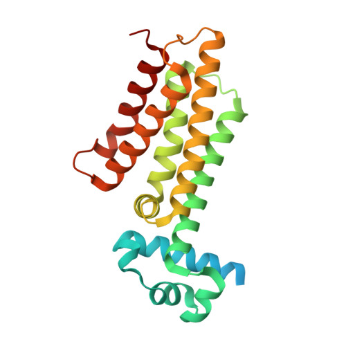
    3O8G, 3O8H

  • PubMed Abstract: 

    In situ click chemistry has been successfully applied to probe the ligand binding domain of EthR, a mycobacterial transcriptional regulator known to control the sensitivity of Mycobacterium tuberculosis to several antibiotics. Specific protein-templated ligands were generated in situ from one azide and six clusters of 10 acetylenic fragments. Comparative X-ray structures of EthR complexed with either clicked ligand BDM14950 or its azide precursor showed ligand-dependent conformational impacts on the protein architecture. This approach revealed two mobile phenylalanine residues that control the access to a previously hidden hydrophobic pocket that can be further exploited for the development of structurally diverse EthR inhibitors. This report shows that protein-directed in situ chemistry allows medicinal chemists to explore the conformational space of a ligand-binding pocket and is thus a valuable tool to guide drug design in the complex path of hit-to-lead processes.

Macromolecule Content 

  • Total Structure Weight: 26.27 kDa 
  • Atom Count: 1,766 
  • Modeled Residue Count: 194 
  • Deposited Residue Count: 236 
  • Unique protein chains: 1

Macromolecules

Find similar proteins by:|  3D Structure
Entity ID: 1
MoleculeChains  Sequence LengthOrganismDetailsImage
TRANSCRIPTIONAL REGULATORY REPRESSOR PROTEIN (TETR-FAMILY) ETHR236Mycobacterium tuberculosisMutation(s): 0 
Gene Names: ethRMT3970Rv3855
UniProt
Find proteins for P9WMC1 (Mycobacterium tuberculosis (strain ATCC 25618 / H37Rv))
Explore P9WMC1 
Go to UniProtKB:  P9WMC1
Entity Groups
Sequence Clusters30% Identity50% Identity70% Identity90% Identity95% Identity100% Identity
UniProt GroupP9WMC1
Sequence Annotations
Expand
Reference Sequence

Small Molecules

Ligands 1 Unique
IDChains Name / Formula / InChI Key2D Diagram3D Interactions
O8G

Query on O8G


Download:Ideal Coordinates CCD File
B [auth A]1-(azidoacetyl)-4-(3-thiophen-2-yl-1,2,4-oxadiazol-5-yl)piperidine
C13 H14 N6 O2 S
RJFYHVFZGWNOBV-UHFFFAOYSA-N

Experimental Data & Validation

Experimental Data

  • Method: X-RAY DIFFRACTION
  • Resolution: 1.90 Å
  • R-Value Free:  0.221 (Depositor), 0.218 (DCC) 
  • R-Value Work:  0.182 (Depositor), 0.181 (DCC) 
  • R-Value Observed: 0.184 (Depositor) 
Space Group: P 41 21 2
Unit Cell:
Length ( Å )Angle ( ˚ )
a = 123.01α = 90
b = 123.01β = 90
c = 33.91γ = 90
Software Package:
Software NamePurpose
MAR345dtbdata collection
CCP4model building
REFMACrefinement
XDSdata reduction
XDSdata scaling
CCP4phasing

Structure Validation

View Full Validation Report


Ligand Structure Quality Assessment 


Entry History 

Deposition Data

Revision History  (Full details and data files)

  • Version 1.0: 2010-09-01
    Type: Initial release
  • Version 1.1: 2011-07-13
    Changes: Version format compliance
  • Version 1.2: 2024-02-21
    Changes: Data collection, Database references, Derived calculations