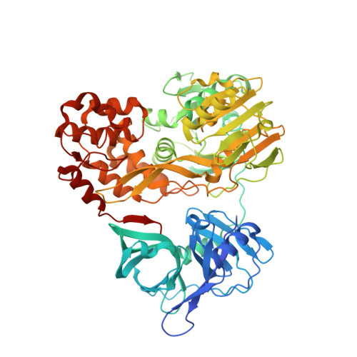
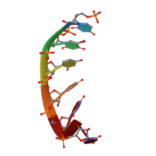
3O8C

Visualizing ATP-dependent RNA Translocation by the NS3 Helicase from HCV


Experimental Data Snapshot

  • Method: X-RAY DIFFRACTION
  • Resolution: 2.00 Å
  • R-Value Free: 0.232 
  • R-Value Work: 0.200 
  • R-Value Observed: 0.200 

Starting Model: experimental
View more details

wwPDB Validation   3D Report Full Report


This is version 1.2 of the entry. See complete history


Literature

Visualizing ATP-Dependent RNA Translocation by the NS3 Helicase from HCV.

Appleby, T.C.Anderson, R.Fedorova, O.Pyle, A.M.Wang, R.Liu, X.Brendza, K.M.Somoza, J.R.

(2011) J Mol Biol 405: 1139-1153

  • DOI: https://doi.org/10.1016/j.jmb.2010.11.034
  • Primary Citation of Related Structures:  
    3O8B, 3O8C, 3O8D, 3O8R

  • PubMed Abstract: 

    The structural mechanism by which nonstructural protein 3 (NS3) from the hepatitis C virus (HCV) translocates along RNA is currently unknown. HCV NS3 is an ATP-dependent motor protein essential for viral replication and a member of the superfamily 2 helicases. Crystallographic analysis using a labeled RNA oligonucleotide allowed us to unambiguously track the positional changes of RNA bound to full-length HCV NS3 during two discrete steps of the ATP hydrolytic cycle. The crystal structures of HCV NS3, NS3 bound to bromine-labeled RNA, and a tertiary complex of NS3 bound to labeled RNA and a non-hydrolyzable ATP analog provide a direct view of how large domain movements resulting from ATP binding and hydrolysis allow the enzyme to translocate along the phosphodiester backbone. While directional translocation of HCV NS3 by a single base pair per ATP hydrolyzed is observed, the 3' end of the RNA does not shift register with respect to a conserved tryptophan residue, supporting a "spring-loading" mechanism that leads to larger steps by the enzyme as it moves along a nucleic acid substrate.


  • Organizational Affiliation

    Department of Structural Chemistry, Gilead Sciences, Inc., 333 Lakeside Drive, Foster City, CA 94404, USA. todd.appleby@gilead.com


Macromolecules

Find similar proteins by:  (by identity cutoff)  |  3D Structure
Entity ID: 1
MoleculeChains Sequence LengthOrganismDetailsImage
HCV NS3 protease/helicaseA,
C [auth B]
666Hepatitis C virus subtype 1bMutation(s): 0 
EC: 3.4.21.98 (PDB Primary Data), 3.6.1.15 (PDB Primary Data), 3.6.4.13 (PDB Primary Data)
UniProt
Find proteins for Q99AU2 (Hepatitis C virus subtype 1b)
Explore Q99AU2 
Go to UniProtKB:  Q99AU2
Entity Groups  
Sequence Clusters30% Identity50% Identity70% Identity90% Identity95% Identity100% Identity
UniProt GroupQ99AU2
Sequence Annotations
Expand
  • Reference Sequence

Find similar nucleic acids by:  Sequence   |   3D Structure  

Entity ID: 2
MoleculeChains LengthOrganismImage
RNA (5'-R(P*UP*(5BU)P*UP*UP*UP*U)-3')B [auth C]6N/A
Sequence Annotations
Expand
  • Reference Sequence
Experimental Data & Validation

Experimental Data

  • Method: X-RAY DIFFRACTION
  • Resolution: 2.00 Å
  • R-Value Free: 0.232 
  • R-Value Work: 0.200 
  • R-Value Observed: 0.200 
  • Space Group: P 21 21 21
Unit Cell:
Length ( Å )Angle ( ˚ )
a = 91.699α = 90
b = 110.166β = 90
c = 142.939γ = 90
Software Package:
Software NamePurpose
SCALEPACKdata scaling
PHENIXrefinement
PDB_EXTRACTdata extraction
BOSdata collection
HKL-2000data reduction
HKL-2000data scaling
EPMRphasing

Structure Validation

View Full Validation Report


Entry History 

Deposition Data

Revision History  (Full details and data files)

  • Version 1.0: 2011-01-05
    Type: Initial release
  • Version 1.1: 2011-07-13
    Changes: Version format compliance
  • Version 1.2: 2023-09-06
    Changes: Data collection, Database references, Derived calculations, Refinement description