3O8R | pdb_00003o8r

Visualizing ATP-dependent RNA Translocation by the NS3 Helicase from HCV


Experimental Data Snapshot

  • Method: X-RAY DIFFRACTION
  • Resolution: 2.30 Å
  • R-Value Free: 
    0.263 (Depositor), 0.253 (DCC) 
  • R-Value Work: 
    0.215 (Depositor), 0.209 (DCC) 
  • R-Value Observed: 
    0.216 (Depositor) 

Starting Model: experimental
View more details

wwPDB Validation 3D Report Full Report

Validation slider image for 3O8R

Ligand Structure Quality Assessment 


This is version 1.2 of the entry. See complete history

Literature

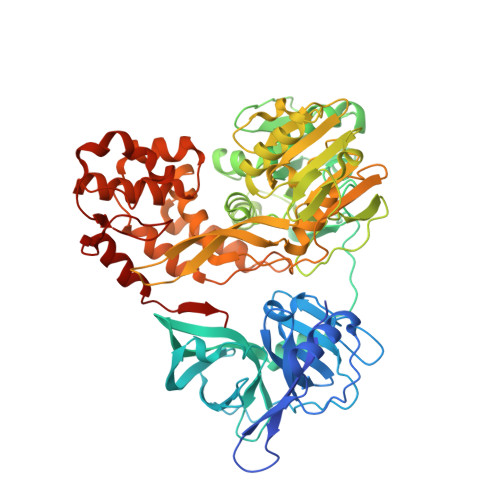
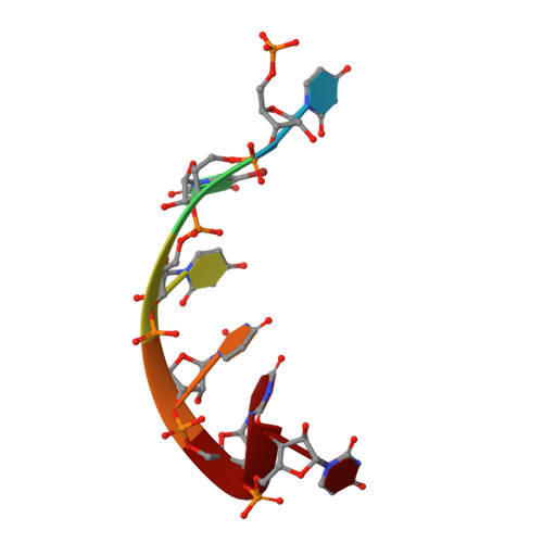
Visualizing ATP-Dependent RNA Translocation by the NS3 Helicase from HCV.

Appleby, T.C.Anderson, R.Fedorova, O.Pyle, A.M.Wang, R.Liu, X.Brendza, K.M.Somoza, J.R.

(2011) J Mol Biology 405: 1139-1153

  • DOI: https://doi.org/10.1016/j.jmb.2010.11.034
  • Primary Citation Related Structures: 
    3O8B, 3O8C, 3O8D, 3O8R

  • PubMed Abstract: 

    The structural mechanism by which nonstructural protein 3 (NS3) from the hepatitis C virus (HCV) translocates along RNA is currently unknown. HCV NS3 is an ATP-dependent motor protein essential for viral replication and a member of the superfamily 2 helicases. Crystallographic analysis using a labeled RNA oligonucleotide allowed us to unambiguously track the positional changes of RNA bound to full-length HCV NS3 during two discrete steps of the ATP hydrolytic cycle. The crystal structures of HCV NS3, NS3 bound to bromine-labeled RNA, and a tertiary complex of NS3 bound to labeled RNA and a non-hydrolyzable ATP analog provide a direct view of how large domain movements resulting from ATP binding and hydrolysis allow the enzyme to translocate along the phosphodiester backbone. While directional translocation of HCV NS3 by a single base pair per ATP hydrolyzed is observed, the 3' end of the RNA does not shift register with respect to a conserved tryptophan residue, supporting a "spring-loading" mechanism that leads to larger steps by the enzyme as it moves along a nucleic acid substrate.


  • Organizational Affiliation
    • Department of Structural Chemistry, Gilead Sciences, Inc., 333 Lakeside Drive, Foster City, CA 94404, USA. todd.appleby@gilead.com

Macromolecule Content 

  • Total Structure Weight: 144.45 kDa 
  • Atom Count: 10,085 
  • Modeled Residue Count: 1,296 
  • Deposited Residue Count: 1,338 
  • Unique protein chains: 1
  • Unique nucleic acid chains: 1

Macromolecules


Find similar proteins by:|  3D Structure
Entity ID: 1
MoleculeChains  Sequence LengthOrganismDetailsImage
HCV NS3 protease/helicaseA,
C [auth B]
666Hepatitis C virus subtype 1bMutation(s): 0 
EC: 3.4.21.98 (PDB Primary Data), 3.6.1.15 (PDB Primary Data), 3.6.4.13 (PDB Primary Data)
UniProt
Find proteins for Q99AU2 (Hepatitis C virus genotype 1b)
Explore Q99AU2 
Go to UniProtKB:  Q99AU2
Entity Groups
Sequence Clusters30% Identity50% Identity70% Identity90% Identity95% Identity100% Identity
UniProt GroupQ99AU2
Sequence Annotations
Expand
Reference Sequence
Find similar nucleic acids by:  Sequence
Entity ID: 2
MoleculeChains LengthOrganismImage
RNA (5'-R(P*UP*(5BU)P*UP*UP*UP*U)-3')B [auth C]6N/A
Sequence Annotations
Expand
Reference Sequence

Small Molecules

Ligands 5 Unique
IDChains Name / Formula / InChI Key2D Diagram3D Interactions
ADP

Query on ADP


Download:Ideal Coordinates CCD File
F [auth A]ADENOSINE-5'-DIPHOSPHATE
C10 H15 N5 O10 P2
XTWYTFMLZFPYCI-KQYNXXCUSA-N
SO4

Query on SO4


Download:Ideal Coordinates CCD File
I [auth B],
J [auth B]
SULFATE ION
O4 S
QAOWNCQODCNURD-UHFFFAOYSA-L
BEF

Query on BEF


Download:Ideal Coordinates CCD File
G [auth A]BERYLLIUM TRIFLUORIDE ION
Be F3
OGIAHMCCNXDTIE-UHFFFAOYSA-K
ZN

Query on ZN


Download:Ideal Coordinates CCD File
D [auth A],
H [auth B]
ZINC ION
Zn
PTFCDOFLOPIGGS-UHFFFAOYSA-N
MG

Query on MG


Download:Ideal Coordinates CCD File
E [auth A]MAGNESIUM ION
Mg
JLVVSXFLKOJNIY-UHFFFAOYSA-N

Experimental Data & Validation

Experimental Data

  • Method: X-RAY DIFFRACTION
  • Resolution: 2.30 Å
  • R-Value Free:  0.263 (Depositor), 0.253 (DCC) 
  • R-Value Work:  0.215 (Depositor), 0.209 (DCC) 
  • R-Value Observed: 0.216 (Depositor) 
Space Group: P 21 21 21
Unit Cell:
Length ( Å )Angle ( ˚ )
a = 91.033α = 90
b = 110.827β = 90
c = 141.979γ = 90
Software Package:
Software NamePurpose
SCALEPACKdata scaling
PHENIXrefinement
PDB_EXTRACTdata extraction
BOSdata collection
HKL-2000data reduction
EPMRphasing

Structure Validation

View Full Validation Report


Ligand Structure Quality Assessment 


Entry History 

Deposition Data

Revision History  (Full details and data files)

  • Version 1.0: 2011-01-05
    Type: Initial release
  • Version 1.1: 2011-07-13
    Changes: Version format compliance
  • Version 1.2: 2023-09-06
    Changes: Data collection, Database references, Derived calculations, Refinement description