3NYK | pdb_00003nyk

The structure of cobalt-substituted pseudoazurin from Alcaligenes faecalis


Experimental Data Snapshot

  • Method: X-RAY DIFFRACTION
  • Resolution: 1.86 Å
  • R-Value Free: 
    0.178 (Depositor), 0.182 (DCC) 
  • R-Value Work: 
    0.150 (Depositor), 0.160 (DCC) 
  • R-Value Observed: 
    0.151 (Depositor) 

wwPDB Validation 3D Report Full Report

Validation slider image for 3NYK

This is version 1.2 of the entry. See complete history

Literature

The crystal structure of cobalt-substituted pseudoazurin from Alcaligenes faecalis.

Gessmann, R.Kyvelidou, C.Papadovasilaki, M.Petratos, K.

(2011) Biopolymers 95: 202-207

  • DOI: https://doi.org/10.1002/bip.21553
  • Primary Citation Related Structures: 
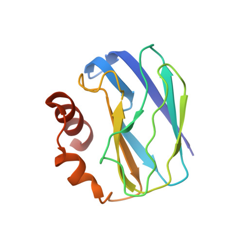
    3NYK

  • PubMed Abstract: 

    The Cu(II) center at the active site of the blue copper protein pseudoazurin from Alcaligenes faecalis has been substituted by Co(II) via denaturing of the protein, chelation and removal of copper by EDTA and refolding of the apo-protein, followed by addition of an aqueous solution of CoCl(2). Sitting drop vapour diffusion experiments produced green hexagonal crystals, which belong to space group P6(5), with unit cell dimensions a = b = 50.03, c = 98.80 Å. Diffraction data, collected at 291 K on a copper rotating anode X-ray source, were phased by the anomalous signal of the cobalt atom. The structure was built automatically, fitted manually and subsequently refined to 1.86 Å resolution. The Co-substituted protein exhibits similar overall geometry to the native structure with copper. Cobalt binds more strongly to the axial Met86-Sδ and retains the tetrahedral arrangement with the four ligand atoms, His40-Nδ(1), Cys78-Sγ, His81-Nδ(1), and 86Met-Sδ, although the structure is less distorted than the native copper protein. The structure reported herein, is the first crystallographic structure of a Co(II)-substituted pseudoazurin.


  • Organizational Affiliation
    • I.M.B.B.-FO.R.T.H., N. Plastira 100, Heraklion 70013, Greece.

Macromolecule Content 

  • Total Structure Weight: 13.44 kDa 
  • Atom Count: 1,080 
  • Modeled Residue Count: 120 
  • Deposited Residue Count: 123 
  • Unique protein chains: 1

Macromolecules

Find similar proteins by:|  3D Structure
Entity ID: 1
MoleculeChains  Sequence LengthOrganismDetailsImage
Pseudoazurin123Alcaligenes faecalisMutation(s): 0 
UniProt
Find proteins for P04377 (Alcaligenes faecalis)
Explore P04377 
Go to UniProtKB:  P04377
Entity Groups
Sequence Clusters30% Identity50% Identity70% Identity90% Identity95% Identity100% Identity
UniProt GroupP04377
Sequence Annotations
Expand
Reference Sequence

Small Molecules

Ligands 1 Unique
IDChains Name / Formula / InChI Key2D Diagram3D Interactions
CO

Query on CO


Download:Ideal Coordinates CCD File
B [auth A]COBALT (II) ION
Co
XLJKHNWPARRRJB-UHFFFAOYSA-N

Experimental Data & Validation

Experimental Data

  • Method: X-RAY DIFFRACTION
  • Resolution: 1.86 Å
  • R-Value Free:  0.178 (Depositor), 0.182 (DCC) 
  • R-Value Work:  0.150 (Depositor), 0.160 (DCC) 
  • R-Value Observed: 0.151 (Depositor) 
Space Group: P 65
Unit Cell:
Length ( Å )Angle ( ˚ )
a = 50.033α = 90
b = 50.033β = 90
c = 98.803γ = 120
Software Package:
Software NamePurpose
Pattersonmodel building
REFMACrefinement
DENZOdata reduction
SCALEPACKdata scaling
Pattersonphasing

Structure Validation

View Full Validation Report


Entry History 

Deposition Data

Revision History  (Full details and data files)

  • Version 1.0: 2010-10-20
    Type: Initial release
  • Version 1.1: 2011-07-13
    Changes: Version format compliance
  • Version 1.2: 2024-02-21
    Changes: Data collection, Database references, Derived calculations