3NGG | pdb_00003ngg

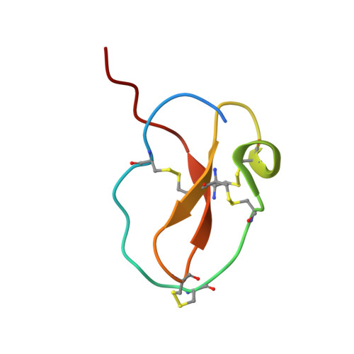
X-ray Structure of Omwaprin


Experimental Data Snapshot

  • Method: X-RAY DIFFRACTION
  • Resolution: 1.33 Å
  • R-Value Free: 
    0.210 (Depositor), 0.232 (DCC) 
  • R-Value Work: 
    0.174 (Depositor) 
  • R-Value Observed: 
    0.176 (Depositor) 

wwPDB Validation 3D Report Full Report

Validation slider image for 3NGG

This is version 1.2 of the entry. See complete history. 

Literature

Determination of the X-ray structure of the snake venom protein omwaprin by total chemical synthesis and racemic protein crystallography.

Banigan, J.R., Mandal, K., Sawaya, M.R., Thammavongsa, V., Hendrickx, A.P., Schneewind, O., Yeates, T.O., Kent, S.B.

(2010) Protein Sci 19: 1840-1849

  • DOI: https://doi.org/10.1002/pro.468
  • Primary Citation Related Structures: 
    3NGG

  • PubMed Abstract: 

    The 50-residue snake venom protein L-omwaprin and its enantiomer D-omwaprin were prepared by total chemical synthesis. Radial diffusion assays were performed against Bacillus megaterium and Bacillus anthracis; both L- and D-omwaprin showed antibacterial activity against B. megaterium. The native protein enantiomer, made of L-amino acids, failed to crystallize readily. However, when a racemic mixture containing equal amounts of L- and D-omwaprin was used, diffraction quality crystals were obtained. The racemic protein sample crystallized in the centrosymmetric space group P2(1)/c and its structure was determined at atomic resolution (1.33 A) by a combination of Patterson and direct methods based on the strong scattering from the sulfur atoms in the eight cysteine residues per protein. Racemic crystallography once again proved to be a valuable method for obtaining crystals of recalcitrant proteins and for determining high-resolution X-ray structures by direct methods.


  • Organizational Affiliation: 
    • Department of Biochemistry and Molecular Biology, The University of Chicago, Chicago, Illinois, USA.

Macromolecule Content 

  • Total Structure Weight: 11.24 kDa 
  • Atom Count: 872 
  • Modeled Residue Count: 94 
  • Deposited Residue Count: 100 
  • Unique protein chains: 1

Macromolecules

Find similar proteins by:|  3D Structure
Entity ID: 1
MoleculeChains  Sequence LengthOrganismDetailsImage
Omwaprin-a
A, B
50Oxyuranus microlepidotusMutation(s): 0 
UniProt
Find proteins for P83952 (Oxyuranus microlepidotus)
Explore P83952 
Go to UniProtKB:  P83952
Entity Groups
Sequence Clusters30% Identity50% Identity70% Identity90% Identity95% Identity100% Identity
UniProt GroupP83952
Sequence Annotations
Expand
Reference Sequence

Experimental Data & Validation

Experimental Data

  • Method: X-RAY DIFFRACTION
  • Resolution: 1.33 Å
  • R-Value Free:  0.210 (Depositor), 0.232 (DCC) 
  • R-Value Work:  0.174 (Depositor) 
  • R-Value Observed: 0.176 (Depositor) 
Space Group: P 1 21/c 1
Unit Cell:
Length ( Å )Angle ( ˚ )
a = 44.09α = 90
b = 46.558β = 92.75
c = 39.65γ = 90
Software Package:
Software NamePurpose
HKL-2000data collection
SHELXDphasing
REFMACrefinement
HKL-2000data reduction
HKL-2000data scaling

Structure Validation

View Full Validation Report


Entry History 

Deposition Data

Revision History  (Full details and data files)

  • Version 1.0: 2010-09-08
    Type: Initial release
  • Version 1.1: 2011-07-13
    Changes: Version format compliance
  • Version 1.2: 2024-10-09
    Changes: Data collection, Database references, Structure summary