3NFZ | pdb_00003nfz

Crystal structure of murine aminoacylase 3 in complex with N-acetyl-L-tyrosine


Experimental Data Snapshot

  • Method: X-RAY DIFFRACTION
  • Resolution: 2.15 Å
  • R-Value Free: 
    0.197 (Depositor), 0.195 (DCC) 
  • R-Value Work: 
    0.166 (Depositor), 0.165 (DCC) 
  • R-Value Observed: 
    0.168 (Depositor) 

Starting Model: experimental
View more details

wwPDB Validation 3D Report Full Report

Validation slider image for 3NFZ

Ligand Structure Quality Assessment 


This is version 1.3 of the entry. See complete history

Literature

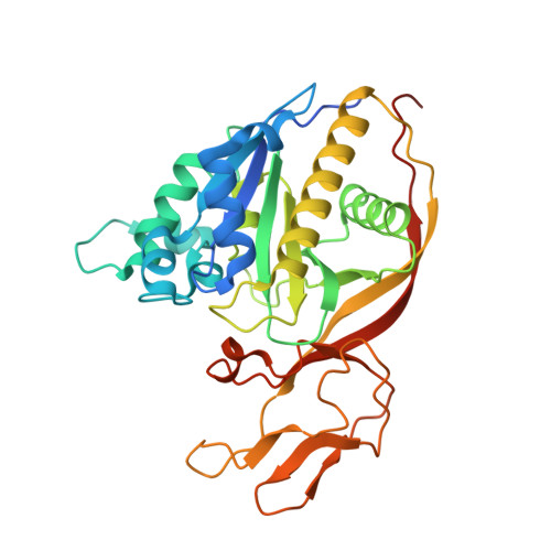
Structures of aminoacylase 3 in complex with acetylated substrates.

Hsieh, J.M.Tsirulnikov, K.Sawaya, M.R.Magilnick, N.Abuladze, N.Kurtz, I.Abramson, J.Pushkin, A.

(2010) Proc Natl Acad Sci U S A 107: 17962-17967

  • DOI: https://doi.org/10.1073/pnas.1006687107
  • Primary Citation Related Structures: 
    3NFZ, 3NH4, 3NH5, 3NH8

  • PubMed Abstract: 

    Trichloroethylene (TCE) is one of the most widespread environmental contaminants, which is metabolized to N-acetyl-S-1,2-dichlorovinyl-L-cysteine (NA-DCVC) before being excreted in the urine. Alternatively, NA-DCVC can be deacetylated by aminoacylase 3 (AA3), an enzyme that is highly expressed in the kidney, liver, and brain. NA-DCVC deacetylation initiates the transformation into toxic products that ultimately causes acute renal failure. AA3 inhibition is therefore a target of interest to prevent TCE induced nephrotoxicity. Here we report the crystal structure of recombinant mouse AA3 (mAA3) in the presence of its acetate byproduct and two substrates: N(α)-acetyl-L-tyrosine and NA-DCVC. These structures, in conjunction with biochemical data, indicated that AA3 mediates substrate specificity through van der Waals interactions providing a dynamic interaction interface, which facilitates a diverse range of substrates.


  • Organizational Affiliation
    • Department of Chemistry and Biochemistry, University of California, Los Angeles, CA 90095, USA.

Macromolecule Content 

  • Total Structure Weight: 36.79 kDa 
  • Atom Count: 2,490 
  • Modeled Residue Count: 305 
  • Deposited Residue Count: 327 
  • Unique protein chains: 1

Macromolecules

Find similar proteins by:|  3D Structure
Entity ID: 1
MoleculeChains  Sequence LengthOrganismDetailsImage
Aspartoacylase-2327Mus musculusMutation(s): 1 
Gene Names: ACY-3Acy3Aspa2
EC: 3.5.1 (PDB Primary Data), 3.5.1.114 (UniProt)
UniProt & NIH Common Fund Data Resources
Find proteins for Q91XE4 (Mus musculus)
Explore Q91XE4 
Go to UniProtKB:  Q91XE4
IMPC:  MGI:1918920
Entity Groups
Sequence Clusters30% Identity50% Identity70% Identity90% Identity95% Identity100% Identity
UniProt GroupQ91XE4
Sequence Annotations
Expand
Reference Sequence

Experimental Data & Validation

Experimental Data

  • Method: X-RAY DIFFRACTION
  • Resolution: 2.15 Å
  • R-Value Free:  0.197 (Depositor), 0.195 (DCC) 
  • R-Value Work:  0.166 (Depositor), 0.165 (DCC) 
  • R-Value Observed: 0.168 (Depositor) 
Space Group: P 62
Unit Cell:
Length ( Å )Angle ( ˚ )
a = 93.498α = 90
b = 93.498β = 90
c = 98.037γ = 120
Software Package:
Software NamePurpose
SCALEPACKdata scaling
PHASERphasing
PHENIXrefinement
PDB_EXTRACTdata extraction
BOSdata collection
DENZOdata reduction

Structure Validation

View Full Validation Report


Ligand Structure Quality Assessment 


Entry History 

Deposition Data

Revision History  (Full details and data files)

  • Version 1.0: 2010-10-20
    Type: Initial release
  • Version 1.1: 2011-07-13
    Changes: Version format compliance
  • Version 1.2: 2024-02-21
    Changes: Data collection, Database references, Derived calculations
  • Version 1.3: 2024-04-03
    Changes: Refinement description