3N10 | pdb_00003n10

Product complex of adenylate cyclase class IV


Experimental Data Snapshot

  • Method: X-RAY DIFFRACTION
  • Resolution: 1.60 Å
  • R-Value Free: 
    0.251 (Depositor), 0.252 (DCC) 
  • R-Value Work: 
    0.218 (Depositor), 0.218 (DCC) 
  • R-Value Observed: 
    0.219 (Depositor) 

Starting Model: experimental
View more details

wwPDB Validation 3D Report Full Report

Validation slider image for 3N10

Ligand Structure Quality Assessment 


This is version 1.4 of the entry. See complete history

Literature

Active-Site Structure of Class IV Adenylyl Cyclase and Transphyletic Mechanism.

Gallagher, D.T.Kim, S.K.Robinson, H.Reddy, P.T.

(2011) J Mol Biology 405: 787-803

  • DOI: https://doi.org/10.1016/j.jmb.2010.11.026
  • Primary Citation Related Structures: 
    3N0Y, 3N0Z, 3N10

  • PubMed Abstract: 

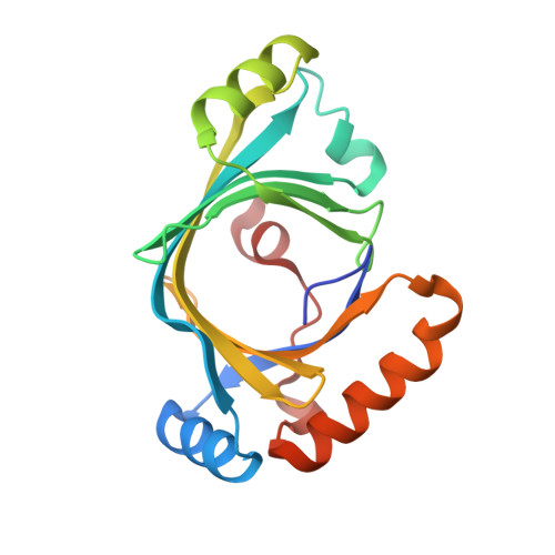
    Adenylyl cyclases (ACs) belonging to three nonhomologous classes (II, III, and IV) have been structurally characterized, enabling a comparison of the mechanisms of cyclic adenosine 3',5'-monophosphate biosynthesis. We report the crystal structures of three active-site complexes for Yersinia pestis class IV AC (AC-IV)-two with substrate analogs and one with product. Mn(2+) binds to all three phosphates, and to Glu12 and Glu136. Electropositive residues Lys14, Arg63, Lys76, Lys111, and Arg113 also form hydrogen bonds to phosphates. The conformation of the analogs is suitable for in-line nucleophilic attack by the ribose O3' on α-phosphate (distance ∼4 Å). In the product complex, a second Mn ion is observed to be coordinated to both ribose 2' oxygen and ribose 3' oxygen. Observation of both metal sites, together with kinetic measurements, provides strong support for a two-cation mechanism. Eleven active-site mutants were also made and kinetically characterized. These findings and comparisons with class II and class III enzymes enable a detailed transphyletic analysis of the AC mechanism. Consistent with its lack of coordination to purine, Y. pestis AC-IV cyclizes both ATP and GTP. As in other classes of AC, the ribose is loosely bound, and as in class III, no base appears to ionize the O3' nucleophile. Different syn/anti conformations suggest that the mechanism involves a conformational transition, and further evidence suggests a role for ribosyl pseudorotation. With resolutions of 1.6-1.7 Å, these are the most detailed active-site ligand complexes for any class of this ubiquitous signaling enzyme.


  • Organizational Affiliation
    • Biochemical Science Division, Chemical Science and Technology Laboratory, National Institute of Standards and Technology, Gaithersburg, MD 20899, USA. gallagher@ibbr.umd.edu

Macromolecule Content 

  • Total Structure Weight: 41.72 kDa 
  • Atom Count: 3,040 
  • Modeled Residue Count: 352 
  • Deposited Residue Count: 358 
  • Unique protein chains: 1

Macromolecules

Find similar proteins by:|  3D Structure
Entity ID: 1
MoleculeChains  Sequence LengthOrganismDetailsImage
Adenylate cyclase 2
A, B
179Yersinia pestisMutation(s): 1 
Gene Names: cyaBy3011YPO1178YP_0959
EC: 4.6.1.1
UniProt
Find proteins for A0A2U2H3Y1 (Yersinia pestis)
Explore A0A2U2H3Y1 
Go to UniProtKB:  A0A2U2H3Y1
Entity Groups
Sequence Clusters30% Identity50% Identity70% Identity90% Identity95% Identity100% Identity
UniProt GroupA0A2U2H3Y1
Sequence Annotations
Expand
Reference Sequence

Small Molecules

Ligands 3 Unique
IDChains Name / Formula / InChI Key2D Diagram3D Interactions
CMP

Query on CMP


Download:Ideal Coordinates CCD File
C [auth A],
G [auth B]
ADENOSINE-3',5'-CYCLIC-MONOPHOSPHATE
C10 H12 N5 O6 P
IVOMOUWHDPKRLL-KQYNXXCUSA-N
SO4

Query on SO4


Download:Ideal Coordinates CCD File
F [auth A],
I [auth B]
SULFATE ION
O4 S
QAOWNCQODCNURD-UHFFFAOYSA-L
MN

Query on MN


Download:Ideal Coordinates CCD File
D [auth A],
E [auth A],
H [auth B]
MANGANESE (II) ION
Mn
WAEMQWOKJMHJLA-UHFFFAOYSA-N

Experimental Data & Validation

Experimental Data

  • Method: X-RAY DIFFRACTION
  • Resolution: 1.60 Å
  • R-Value Free:  0.251 (Depositor), 0.252 (DCC) 
  • R-Value Work:  0.218 (Depositor), 0.218 (DCC) 
  • R-Value Observed: 0.219 (Depositor) 
Space Group: P 1 21 1
Unit Cell:
Length ( Å )Angle ( ˚ )
a = 64.477α = 90
b = 38.124β = 98.71
c = 80.658γ = 90
Software Package:
Software NamePurpose
DENZOdata reduction
SCALEPACKdata scaling
PHASERphasing
REFMACrefinement
PDB_EXTRACTdata extraction
HKL-3000data collection

Structure Validation

View Full Validation Report


Ligand Structure Quality Assessment 


Entry History 

Deposition Data

Revision History  (Full details and data files)

  • Version 1.0: 2010-06-23
    Type: Initial release
  • Version 1.1: 2011-07-13
    Changes: Version format compliance
  • Version 1.2: 2017-03-22
    Changes: Source and taxonomy
  • Version 1.3: 2017-11-08
    Changes: Refinement description
  • Version 1.4: 2023-09-06
    Changes: Data collection, Database references, Derived calculations, Refinement description