3KXA | pdb_00003kxa

Crystal Structure of NGO0477 from Neisseria gonorrhoeae


Experimental Data Snapshot

  • Method: X-RAY DIFFRACTION
  • Resolution: 2.80 Å
  • R-Value Free: 
    0.280 (Depositor), 0.232 (DCC) 
  • R-Value Work: 
    0.230 (Depositor), 0.238 (DCC) 
  • R-Value Observed: 
    0.232 (Depositor) 

wwPDB Validation 3D Report Full Report

Validation slider image for 3KXA

This is version 1.2 of the entry. See complete history

Literature

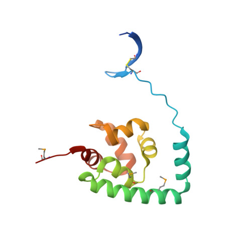
The crystal structure of NGO0477 from Neisseria gonorrhoeae reveals a novel protein fold incorporating a helix-turn-helix motif.

Ren, J.Sainsbury, S.Nettleship, J.E.Saunders, N.J.Owens, R.J.

(2010) Proteins 78: 1798-1802

Macromolecule Content 

  • Total Structure Weight: 64.97 kDa 
  • Atom Count: 4,160 
  • Modeled Residue Count: 522 
  • Deposited Residue Count: 564 
  • Unique protein chains: 1

Macromolecules

Find similar proteins by:|  3D Structure
Entity ID: 1
MoleculeChains  Sequence LengthOrganismDetailsImage
Putative uncharacterized protein
A, B, C, D
141Neisseria gonorrhoeae FA 1090Mutation(s): 0 
Gene Names: NGO0477
UniProt
Find proteins for Q5F9C2 (Neisseria gonorrhoeae (strain ATCC 700825 / FA 1090))
Explore Q5F9C2 
Go to UniProtKB:  Q5F9C2
Entity Groups
Sequence Clusters30% Identity50% Identity70% Identity90% Identity95% Identity100% Identity
UniProt GroupQ5F9C2
Sequence Annotations
Expand
Reference Sequence

Small Molecules

Ligands 3 Unique
IDChains Name / Formula / InChI Key2D Diagram3D Interactions
ASN

Query on ASN


Download:Ideal Coordinates CCD File
E [auth A],
QA [auth D],
V [auth B],
Y [auth C]
ASPARAGINE
C4 H8 N2 O3
DCXYFEDJOCDNAF-REOHCLBHSA-N
CL

Query on CL


Download:Ideal Coordinates CCD File
F [auth A]
G [auth A]
H [auth A]
I [auth A]
J [auth A]
F [auth A],
G [auth A],
H [auth A],
I [auth A],
J [auth A],
KA [auth C],
LA [auth C],
MA [auth C],
NA [auth C],
OA [auth C],
PA [auth C],
W [auth B],
X [auth B]
CHLORIDE ION
Cl
VEXZGXHMUGYJMC-UHFFFAOYSA-M
UNX

Query on UNX


Download:Ideal Coordinates CCD File
AA [auth C]
BA [auth C]
CA [auth C]
DA [auth C]
EA [auth C]
AA [auth C],
BA [auth C],
CA [auth C],
DA [auth C],
EA [auth C],
FA [auth C],
GA [auth C],
HA [auth C],
IA [auth C],
JA [auth C],
K [auth A],
L [auth A],
M [auth A],
N [auth A],
O [auth A],
P [auth A],
Q [auth A],
R [auth A],
S [auth A],
T [auth A],
U [auth A],
Z [auth C]
UNKNOWN ATOM OR ION
X
Modified Residues  1 Unique
IDChains TypeFormula2D DiagramParent
MSE
Query on MSE
A, B, C, D
L-PEPTIDE LINKINGC5 H11 N O2 SeMET

Experimental Data & Validation

Experimental Data

  • Method: X-RAY DIFFRACTION
  • Resolution: 2.80 Å
  • R-Value Free:  0.280 (Depositor), 0.232 (DCC) 
  • R-Value Work:  0.230 (Depositor), 0.238 (DCC) 
  • R-Value Observed: 0.232 (Depositor) 
Space Group: H 3
Unit Cell:
Length ( Å )Angle ( ˚ )
a = 124.823α = 90
b = 124.823β = 90
c = 137.577γ = 120
Software Package:
Software NamePurpose
SOLVEphasing
REFMACrefinement
HKL-2000data reduction
HKL-2000data scaling

Structure Validation

View Full Validation Report


Entry History 

Deposition Data

Revision History  (Full details and data files)

  • Version 1.0: 2010-01-19
    Type: Initial release
  • Version 1.1: 2011-07-13
    Changes: Advisory, Refinement description, Version format compliance
  • Version 1.2: 2024-10-30
    Changes: Data collection, Database references, Derived calculations, Refinement description, Structure summary