3KPH | pdb_00003kph

Crystal structure of Mycoplasma arthritidis-derived mitogen


Experimental Data Snapshot

  • Method: X-RAY DIFFRACTION
  • Resolution: 2.80 Å
  • R-Value Free: 
    0.299 (Depositor), 0.292 (DCC) 
  • R-Value Work: 
    0.253 (Depositor), 0.251 (DCC) 
  • R-Value Observed: 
    0.253 (Depositor) 

Starting Model: experimental
View more details

wwPDB Validation 3D Report Full Report

Validation slider image for 3KPH

This is version 1.3 of the entry. See complete history

Literature

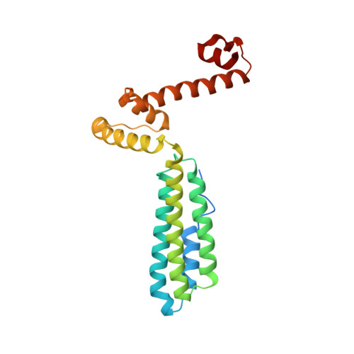
Crystal Structure of the Mycoplasma arthritidis-Derived Mitogen in Apo Form Reveals a 3D Domain-Swapped Dimer.

Liu, L.Li, Z.Guo, Y.Vanvranken, S.J.Mourad, W.Li, H.

(2010) J Mol Biology 399: 367-376

  • DOI: https://doi.org/10.1016/j.jmb.2010.04.030
  • Primary Citation Related Structures: 
    3KPH

  • PubMed Abstract: 

    Mycoplasma arthritidis-derived mitogen (MAM) is a superantigen that can activate large fractions of T cells bearing particular Vbeta elements of T cell receptor. Here, we report the crystal structure of a MAM mutant K201A in apo form (unliganded) at 2.8-A resolutions. We also partially refined the crystal structures of the MAM wild type and another MAM mutant L50A in apo forms at low resolutions. Unexpectedly, the structures of these apo MAM molecules display a three-dimensional domain-swapped dimer. The entire C-terminal domains of these MAM molecules are involved in the domain swapping. Functional analyses demonstrated that the K201A and L50A mutants do not show altered ability to bind to their host receptors and that they stimulate the activation of T cells as efficiently as does the wild type. Structural comparisons indicated that the "reconstituted" MAM monomer from the domain-swapped dimer displays large differences at the hinge regions from the MAM(wt) molecule in the receptor-bound form. Further comparison indicated that MAM has a flexible N-terminal loop, implying that conformational changes could occur upon receptor binding.


  • Organizational Affiliation
    • Wadsworth Center, New York State Department of Health, 120 New Scotland Ave, Albany, NY 12208, USA.

Macromolecule Content 

  • Total Structure Weight: 51.43 kDa 
  • Atom Count: 3,336 
  • Modeled Residue Count: 390 
  • Deposited Residue Count: 434 
  • Unique protein chains: 1

Macromolecules

Find similar proteins by:|  3D Structure
Entity ID: 1
MoleculeChains  Sequence LengthOrganismDetailsImage
Superantigen
A, B
217Metamycoplasma arthritidisMutation(s): 1 
UniProt
Find proteins for Q48898 (Metamycoplasma arthritidis)
Explore Q48898 
Go to UniProtKB:  Q48898
Entity Groups
Sequence Clusters30% Identity50% Identity70% Identity90% Identity95% Identity100% Identity
UniProt GroupQ48898
Sequence Annotations
Expand
Reference Sequence

Experimental Data & Validation

Experimental Data

  • Method: X-RAY DIFFRACTION
  • Resolution: 2.80 Å
  • R-Value Free:  0.299 (Depositor), 0.292 (DCC) 
  • R-Value Work:  0.253 (Depositor), 0.251 (DCC) 
  • R-Value Observed: 0.253 (Depositor) 
Space Group: P 41 3 2
Unit Cell:
Length ( Å )Angle ( ˚ )
a = 181.865α = 90
b = 181.865β = 90
c = 181.865γ = 90
Software Package:
Software NamePurpose
CNSrefinement
PDB_EXTRACTdata extraction
ADSCdata collection
d*TREKdata reduction
d*TREKdata scaling
PHASERphasing

Structure Validation

View Full Validation Report


Entry History 

Deposition Data

Revision History  (Full details and data files)

  • Version 1.0: 2010-05-12
    Type: Initial release
  • Version 1.1: 2011-07-13
    Changes: Version format compliance
  • Version 1.2: 2021-10-13
    Changes: Database references, Derived calculations
  • Version 1.3: 2023-09-06
    Changes: Data collection, Refinement description