3KHE | pdb_00003khe

Crystal structure of the calcium-loaded calmodulin-like domain of the CDPK, 541.m00134 from toxoplasma gondii


Experimental Data Snapshot

  • Method: X-RAY DIFFRACTION
  • Resolution: 1.95 Å
  • R-Value Free: 
    0.277 (Depositor), 0.272 (DCC) 
  • R-Value Work: 
    0.201 (Depositor) 
  • R-Value Observed: 
    0.204 (Depositor) 

wwPDB Validation 3D Report Full Report

Validation slider image for 3KHE

This is version 1.3 of the entry. See complete history

Literature

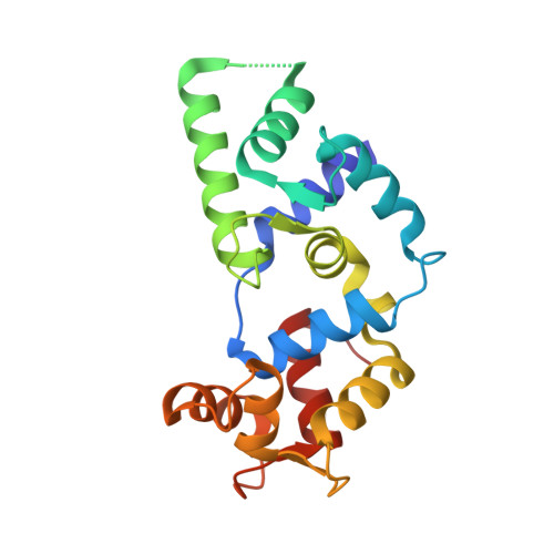
Structures of parasitic CDPK domains point to a common mechanism of activation.

Wernimont, A.K.Amani, M.Qiu, W.Pizarro, J.C.Artz, J.D.Lin, Y.H.Lew, J.Hutchinson, A.Hui, R.

(2011) Proteins 79: 803-820

  • DOI: https://doi.org/10.1002/prot.22919
  • Primary Citation Related Structures: 
    3K21, 3KHE

  • PubMed Abstract: 

    We recently determined the first structures of inactivated and calcium-activated calcium-dependent protein kinases (CDPKs) from Apicomplexa. Calcium binding triggered a large conformational change that constituted a new mechanism in calcium signaling and a novel EF-hand fold (CAD, for CDPK activation domain). Thus we set out to determine if this mechanism was universal to all CDPKs. We solved additional CDPK structures, including one from the species Plasmodium. We highlight the similarities in sequence and structure across apicomplexan and plant CDPKs, and strengthen our observations that this novel mechanism could be universal to canonical CDPKs. Our new structures demonstrate more detailed steps in the mechanism of calcium activation and possible key players in regulation. Residues involved in making the largest conformational change are the most conserved across Apicomplexa, leading us to propose that the mechanism is indeed conserved. CpCDPK3_CAD and PfCDPK_CAD were captured at a possible intermediate conformation, lending insight into the order of activation steps. PfCDPK3_CAD adopts an activated fold, despite having an inactive EF-hand sequence in the N-terminal lobe. We propose that for most apicomplexan CDPKs, the mode of activation will be similar to that seen in our structures, while specific regulation of the inactive and active forms will require further investigation.


  • Organizational Affiliation
    • Structural Genomics Consortium, University of Toronto, Toronto, Ontario, Canada M5G 1L7. amy.wernimont@utoronto.ca

Macromolecule Content 

  • Total Structure Weight: 44.35 kDa 
  • Atom Count: 3,541 
  • Modeled Residue Count: 375 
  • Deposited Residue Count: 382 
  • Unique protein chains: 1

Macromolecules

Find similar proteins by:|  3D Structure
Entity ID: 1
MoleculeChains  Sequence LengthOrganismDetailsImage
Calmodulin-like domain protein kinase isoform 3
A, B
191Toxoplasma gondiiMutation(s): 0 
Gene Names: TGME49_105860
EC: 2.7.11.1
UniProt
Find proteins for Q3HNM6 (Toxoplasma gondii)
Explore Q3HNM6 
Go to UniProtKB:  Q3HNM6
Entity Groups
Sequence Clusters30% Identity50% Identity70% Identity90% Identity95% Identity100% Identity
UniProt GroupQ3HNM6
Sequence Annotations
Expand
Reference Sequence

Small Molecules

Ligands 3 Unique
IDChains Name / Formula / InChI Key2D Diagram3D Interactions
GOL

Query on GOL


Download:Ideal Coordinates CCD File
L [auth B],
O [auth B],
P [auth B]
GLYCEROL
C3 H8 O3
PEDCQBHIVMGVHV-UHFFFAOYSA-N
CA

Query on CA


Download:Ideal Coordinates CCD File
C [auth A]
D [auth A]
E [auth A]
F [auth A]
H [auth B]
C [auth A],
D [auth A],
E [auth A],
F [auth A],
H [auth B],
I [auth B],
J [auth B],
K [auth B]
CALCIUM ION
Ca
BHPQYMZQTOCNFJ-UHFFFAOYSA-N
MG

Query on MG


Download:Ideal Coordinates CCD File
G [auth A],
M [auth B],
N [auth B]
MAGNESIUM ION
Mg
JLVVSXFLKOJNIY-UHFFFAOYSA-N

Experimental Data & Validation

Experimental Data

  • Method: X-RAY DIFFRACTION
  • Resolution: 1.95 Å
  • R-Value Free:  0.277 (Depositor), 0.272 (DCC) 
  • R-Value Work:  0.201 (Depositor) 
  • R-Value Observed: 0.204 (Depositor) 
Space Group: P 21 21 21
Unit Cell:
Length ( Å )Angle ( ˚ )
a = 45.838α = 90
b = 101.015β = 90
c = 107.87γ = 90
Software Package:
Software NamePurpose
DENZOdata reduction
SCALEPACKdata scaling
REFMACrefinement
PDB_EXTRACTdata extraction
JBluIce-EPICSdata collection
BALBESphasing

Structure Validation

View Full Validation Report


Entry History 

Revision History  (Full details and data files)

  • Version 1.0: 2010-01-19
    Type: Initial release
  • Version 1.1: 2011-07-13
    Changes: Version format compliance
  • Version 1.2: 2017-11-01
    Changes: Refinement description
  • Version 1.3: 2024-02-21
    Changes: Data collection, Database references, Derived calculations