3K3C | pdb_00003k3c

The N-terminal PAS domain crystal structure of Rv1364c from Mycobacterium tuberculosis at 1.62


Experimental Data Snapshot

  • Method: X-RAY DIFFRACTION
  • Resolution: 1.62 Å
  • R-Value Free: 
    0.212 (Depositor), 0.211 (DCC) 
  • R-Value Work: 
    0.181 (Depositor) 
  • R-Value Observed: 
    0.183 (Depositor) 

wwPDB Validation 3D Report Full Report

Validation slider image for 3K3C

Ligand Structure Quality Assessment 


This is version 1.2 of the entry. See complete history

Literature

The N-terminal PAS domain crystal structure of Rv1364c from Mycobacterium tuberculosis at 1.62

King-Scott, J.Tucker, P.A.

To be published.

Macromolecule Content 

  • Total Structure Weight: 73.81 kDa 
  • Atom Count: 5,976 
  • Modeled Residue Count: 604 
  • Deposited Residue Count: 632 
  • Unique protein chains: 1

Macromolecules

Find similar proteins by:|  3D Structure
Entity ID: 1
MoleculeChains  Sequence LengthOrganismDetailsImage
Protein Rv1364c/MT1410
A, B, C, D
158Mycobacterium tuberculosisMutation(s): 0 
Gene Names: MT1410MTCY02B10.28cRv1364c
EC: 2.7.11.1 (UniProt), 3.1.3.16 (UniProt)
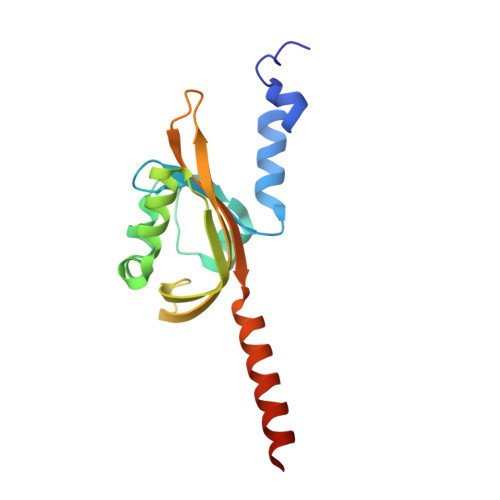
UniProt
Find proteins for P9WLZ7 (Mycobacterium tuberculosis (strain ATCC 25618 / H37Rv))
Explore P9WLZ7 
Go to UniProtKB:  P9WLZ7
Entity Groups
Sequence Clusters30% Identity50% Identity70% Identity90% Identity95% Identity100% Identity
UniProt GroupP9WLZ7
Sequence Annotations
Expand
Reference Sequence

Small Molecules

Ligands 3 Unique
IDChains Name / Formula / InChI Key2D Diagram3D Interactions
PLM

Query on PLM


Download:Ideal Coordinates CCD File
E [auth A],
L [auth B]
PALMITIC ACID
C16 H32 O2
IPCSVZSSVZVIGE-UHFFFAOYSA-N
SO4

Query on SO4


Download:Ideal Coordinates CCD File
F [auth A]
M [auth B]
S [auth C]
T [auth C]
U [auth D]
F [auth A],
M [auth B],
S [auth C],
T [auth C],
U [auth D],
V [auth D]
SULFATE ION
O4 S
QAOWNCQODCNURD-UHFFFAOYSA-L
GOL

Query on GOL


Download:Ideal Coordinates CCD File
G [auth A]
H [auth A]
I [auth A]
J [auth A]
K [auth A]
G [auth A],
H [auth A],
I [auth A],
J [auth A],
K [auth A],
N [auth B],
O [auth B],
P [auth B],
Q [auth B],
R [auth B],
W [auth D]
GLYCEROL
C3 H8 O3
PEDCQBHIVMGVHV-UHFFFAOYSA-N

Experimental Data & Validation

Experimental Data

  • Method: X-RAY DIFFRACTION
  • Resolution: 1.62 Å
  • R-Value Free:  0.212 (Depositor), 0.211 (DCC) 
  • R-Value Work:  0.181 (Depositor) 
  • R-Value Observed: 0.183 (Depositor) 
Space Group: C 1 2 1
Unit Cell:
Length ( Å )Angle ( ˚ )
a = 209.069α = 90
b = 63.823β = 89.95
c = 61.305γ = 90
Software Package:
Software NamePurpose
DENZOdata reduction
Auto-Rickshawphasing
REFMACrefinement
SCALEPACKdata scaling

Structure Validation

View Full Validation Report


Ligand Structure Quality Assessment 


Entry History 

Deposition Data

Revision History  (Full details and data files)

  • Version 1.0: 2010-09-29
    Type: Initial release
  • Version 1.1: 2011-07-13
    Changes: Version format compliance
  • Version 1.2: 2024-02-21
    Changes: Data collection, Database references, Derived calculations, Refinement description