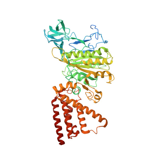
3I72

Structural characterization for the nucleotide binding ability of subunit A with SO4 of the A1AO ATP synthase


Experimental Data Snapshot

  • Method: X-RAY DIFFRACTION
  • Resolution: 2.47 Å
  • R-Value Free: 0.253 
  • R-Value Work: 0.217 
  • R-Value Observed: 0.219 

Starting Model: experimental
View more details

wwPDB Validation   3D Report Full Report


This is version 1.6 of the entry. See complete history


Literature

Nucleotide binding states of subunit A of the A-ATP synthase and the implication of P-loop switch in evolution.

Kumar, A.Manimekalai, M.S.Balakrishna, A.M.Jeyakanthan, J.Gruber, G.

(2010) J Mol Biol 396: 301-320

  • DOI: https://doi.org/10.1016/j.jmb.2009.11.046
  • Primary Citation of Related Structures:  
    3I4L, 3I72, 3I73, 3IKJ

  • PubMed Abstract: 

    The crystal structures of the nucleotide-empty (A(E)), 5'-adenylyl-beta,gamma-imidodiphosphate (A(PNP))-bound, and ADP (A(DP))-bound forms of the catalytic A subunit of the energy producer A(1)A(O) ATP synthase from Pyrococcus horikoshii OT3 have been solved at 2.47 A and 2.4 A resolutions. The structures provide novel features of nucleotide binding and depict the residues involved in the catalysis of the A subunit. In the A(E) form, the phosphate analog SO(4)(2-) binds, via a water molecule, to the phosphate binding loop (P-loop) residue Ser238, which is also involved in the phosphate binding of ADP and 5'-adenylyl-beta,gamma-imidodiphosphate. Together with amino acids Gly234 and Phe236, the serine residue stabilizes the arched P-loop conformation of subunit A, as shown by the 2.4-A structure of the mutant protein S238A in which the P-loop flips into a relaxed state, comparable to the one in catalytic beta subunits of F(1)F(O) ATP synthases. Superposition of the existing P-loop structures of ATPases emphasizes the unique P-loop in subunit A, which is also discussed in the light of an evolutionary P-loop switch in related A(1)A(O) ATP synthases, F(1)F(O) ATP synthases, and vacuolar ATPases and implicates diverse catalytic mechanisms inside these biological motors.


  • Organizational Affiliation

    School of Biological Sciences, Nanyang Technological University, Singapore, Republic of Singapore.


Macromolecules
Find similar proteins by:  (by identity cutoff)  |  3D Structure
Entity ID: 1
MoleculeChains Sequence LengthOrganismDetailsImage
A-TYPE ATP SYNTHASE CATALYTIC SUBUNIT A588Pyrococcus horikoshii OT3Mutation(s): 1 
Gene Names: atpAPH1975
EC: 3.6.3.14 (PDB Primary Data), 7.1.2.2 (UniProt)
UniProt
Find proteins for O57728 (Pyrococcus horikoshii (strain ATCC 700860 / DSM 12428 / JCM 9974 / NBRC 100139 / OT-3))
Explore O57728 
Go to UniProtKB:  O57728
Entity Groups  
Sequence Clusters30% Identity50% Identity70% Identity90% Identity95% Identity100% Identity
UniProt GroupO57728
Sequence Annotations
Expand
  • Reference Sequence
Small Molecules
Ligands 4 Unique
IDChains Name / Formula / InChI Key2D Diagram3D Interactions
TRS
Query on TRS

Download Ideal Coordinates CCD File 
C [auth A]2-AMINO-2-HYDROXYMETHYL-PROPANE-1,3-DIOL
C4 H12 N O3
LENZDBCJOHFCAS-UHFFFAOYSA-O
MPD
Query on MPD

Download Ideal Coordinates CCD File 
E [auth A],
F [auth A],
G [auth A]
(4S)-2-METHYL-2,4-PENTANEDIOL
C6 H14 O2
SVTBMSDMJJWYQN-YFKPBYRVSA-N
SO4
Query on SO4

Download Ideal Coordinates CCD File 
B [auth A]SULFATE ION
O4 S
QAOWNCQODCNURD-UHFFFAOYSA-L
ACY
Query on ACY

Download Ideal Coordinates CCD File 
D [auth A]ACETIC ACID
C2 H4 O2
QTBSBXVTEAMEQO-UHFFFAOYSA-N
Experimental Data & Validation

Experimental Data

  • Method: X-RAY DIFFRACTION
  • Resolution: 2.47 Å
  • R-Value Free: 0.253 
  • R-Value Work: 0.217 
  • R-Value Observed: 0.219 
  • Space Group: P 43 21 2
Unit Cell:
Length ( Å )Angle ( ˚ )
a = 128.248α = 90
b = 128.248β = 90
c = 104.676γ = 90
Software Package:
Software NamePurpose
DENZOdata reduction
SCALEPACKdata scaling
MOLREPphasing
REFMACrefinement
PDB_EXTRACTdata extraction
HKL-2000data collection
HKL-2000data reduction
HKL-2000data scaling

Structure Validation

View Full Validation Report


Entry History 

Deposition Data

Revision History  (Full details and data files)

  • Version 1.0: 2010-01-12
    Type: Initial release
  • Version 1.1: 2011-07-13
    Changes: Version format compliance
  • Version 1.2: 2013-11-13
    Changes: Database references
  • Version 1.3: 2017-08-23
    Changes: Refinement description, Source and taxonomy
  • Version 1.4: 2017-11-01
    Changes: Refinement description
  • Version 1.5: 2021-11-10
    Changes: Database references, Derived calculations
  • Version 1.6: 2023-11-01
    Changes: Data collection, Refinement description