3I73 | pdb_00003i73

Structural characterization for the nucleotide binding ability of subunit A with ADP of the A1AO ATP synthase


Experimental Data Snapshot

  • Method: X-RAY DIFFRACTION
  • Resolution: 2.40 Å
  • R-Value Free: 
    0.258 (Depositor), 0.245 (DCC) 
  • R-Value Work: 
    0.225 (Depositor), 0.230 (DCC) 
  • R-Value Observed: 
    0.227 (Depositor) 

Starting Model: experimental
View more details

wwPDB Validation 3D Report Full Report

Validation slider image for 3I73

Ligand Structure Quality Assessment 


This is version 1.6 of the entry. See complete history

Literature

Nucleotide binding states of subunit A of the A-ATP synthase and the implication of P-loop switch in evolution.

Kumar, A.Manimekalai, M.S.Balakrishna, A.M.Jeyakanthan, J.Gruber, G.

(2010) J Mol Biology 396: 301-320

  • DOI: https://doi.org/10.1016/j.jmb.2009.11.046
  • Primary Citation Related Structures: 
    3I4L, 3I72, 3I73, 3IKJ

  • PubMed Abstract: 

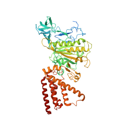
    The crystal structures of the nucleotide-empty (A(E)), 5'-adenylyl-beta,gamma-imidodiphosphate (A(PNP))-bound, and ADP (A(DP))-bound forms of the catalytic A subunit of the energy producer A(1)A(O) ATP synthase from Pyrococcus horikoshii OT3 have been solved at 2.47 A and 2.4 A resolutions. The structures provide novel features of nucleotide binding and depict the residues involved in the catalysis of the A subunit. In the A(E) form, the phosphate analog SO(4)(2-) binds, via a water molecule, to the phosphate binding loop (P-loop) residue Ser238, which is also involved in the phosphate binding of ADP and 5'-adenylyl-beta,gamma-imidodiphosphate. Together with amino acids Gly234 and Phe236, the serine residue stabilizes the arched P-loop conformation of subunit A, as shown by the 2.4-A structure of the mutant protein S238A in which the P-loop flips into a relaxed state, comparable to the one in catalytic beta subunits of F(1)F(O) ATP synthases. Superposition of the existing P-loop structures of ATPases emphasizes the unique P-loop in subunit A, which is also discussed in the light of an evolutionary P-loop switch in related A(1)A(O) ATP synthases, F(1)F(O) ATP synthases, and vacuolar ATPases and implicates diverse catalytic mechanisms inside these biological motors.


  • Organizational Affiliation
    • School of Biological Sciences, Nanyang Technological University, Singapore, Republic of Singapore.

Macromolecule Content 

  • Total Structure Weight: 66.74 kDa 
  • Atom Count: 4,408 
  • Modeled Residue Count: 512 
  • Deposited Residue Count: 588 
  • Unique protein chains: 1

Macromolecules

Find similar proteins by:|  3D Structure
Entity ID: 1
MoleculeChains  Sequence LengthOrganismDetailsImage
A-TYPE ATP SYNTHASE CATALYTIC SUBUNIT A588Pyrococcus horikoshii OT3Mutation(s): 1 
EC: 3.6.3.14 (PDB Primary Data), 7.1.2.2 (UniProt)
UniProt
Find proteins for O57728 (Pyrococcus horikoshii (strain ATCC 700860 / DSM 12428 / JCM 9974 / NBRC 100139 / OT-3))
Explore O57728 
Go to UniProtKB:  O57728
Entity Groups
Sequence Clusters30% Identity50% Identity70% Identity90% Identity95% Identity100% Identity
UniProt GroupO57728
Sequence Annotations
Expand
Reference Sequence

Small Molecules

Ligands 4 Unique
IDChains Name / Formula / InChI Key2D Diagram3D Interactions
ADP

Query on ADP


Download:Ideal Coordinates CCD File
B [auth A]ADENOSINE-5'-DIPHOSPHATE
C10 H15 N5 O10 P2
XTWYTFMLZFPYCI-KQYNXXCUSA-N
TRS

Query on TRS


Download:Ideal Coordinates CCD File
F [auth A]2-AMINO-2-HYDROXYMETHYL-PROPANE-1,3-DIOL
C4 H12 N O3
LENZDBCJOHFCAS-UHFFFAOYSA-O
MPD

Query on MPD


Download:Ideal Coordinates CCD File
C [auth A],
D [auth A],
E [auth A]
(4S)-2-METHYL-2,4-PENTANEDIOL
C6 H14 O2
SVTBMSDMJJWYQN-YFKPBYRVSA-N
ACY

Query on ACY


Download:Ideal Coordinates CCD File
G [auth A]ACETIC ACID
C2 H4 O2
QTBSBXVTEAMEQO-UHFFFAOYSA-N

Experimental Data & Validation

Experimental Data

  • Method: X-RAY DIFFRACTION
  • Resolution: 2.40 Å
  • R-Value Free:  0.258 (Depositor), 0.245 (DCC) 
  • R-Value Work:  0.225 (Depositor), 0.230 (DCC) 
  • R-Value Observed: 0.227 (Depositor) 
Space Group: P 43 21 2
Unit Cell:
Length ( Å )Angle ( ˚ )
a = 128.472α = 90
b = 128.472β = 90
c = 104.991γ = 90
Software Package:
Software NamePurpose
DENZOdata reduction
SCALEPACKdata scaling
MOLREPphasing
REFMACrefinement
PDB_EXTRACTdata extraction
HKL-2000data collection

Structure Validation

View Full Validation Report


Ligand Structure Quality Assessment 


Entry History 

Deposition Data

Revision History  (Full details and data files)

  • Version 1.0: 2010-01-12
    Type: Initial release
  • Version 1.1: 2011-07-13
    Changes: Version format compliance
  • Version 1.2: 2013-11-13
    Changes: Database references
  • Version 1.3: 2017-08-23
    Changes: Refinement description, Source and taxonomy
  • Version 1.4: 2017-11-01
    Changes: Refinement description
  • Version 1.5: 2021-11-10
    Changes: Database references, Derived calculations
  • Version 1.6: 2023-11-01
    Changes: Data collection, Refinement description