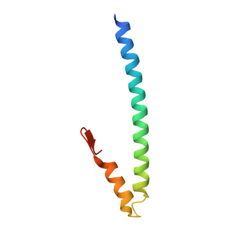
3GJO

Crystal structure of human EB1 in complex with microtubule Tip localization signal peptide of MACF


Experimental Data Snapshot

  • Method: X-RAY DIFFRACTION
  • Resolution: 2.50 Å
  • R-Value Free: 0.255 
  • R-Value Work: 0.213 
  • R-Value Observed: 0.215 

Starting Model: experimental
View more details

wwPDB Validation   3D Report Full Report


This is version 1.3 of the entry. See complete history


Literature

An EB1-binding motif acts as a microtubule tip localization signal

Honnappa, S.Gouveia, S.M.Weisbrich, A.Damberger, F.F.Bhavesh, N.S.Jawhari, H.Grigoriev, I.van Rijssel, F.J.A.Buey, R.M.Lawera, A.Jelesarov, I.Winkler, F.K.Wuthrich, K.Akhmanova, A.Steinmetz, M.O.

(2009) Cell 138: 366-376

  • DOI: https://doi.org/10.1016/j.cell.2009.04.065
  • Primary Citation of Related Structures:  
    3GJO

  • PubMed Abstract: 

    Microtubules are filamentous polymers essential for cell viability. Microtubule plus-end tracking proteins (+TIPs) associate with growing microtubule plus ends and control microtubule dynamics and interactions with different cellular structures during cell division, migration, and morphogenesis. EB1 and its homologs are highly conserved proteins that play an important role in the targeting of +TIPs to microtubule ends, but the underlying molecular mechanism remains elusive. By using live cell experiments and in vitro reconstitution assays, we demonstrate that a short polypeptide motif, Ser-x-Ile-Pro (SxIP), is used by numerous +TIPs, including the tumor suppressor APC, the transmembrane protein STIM1, and the kinesin MCAK, for localization to microtubule tips in an EB1-dependent manner. Structural and biochemical data reveal the molecular basis of the EB1-SxIP interaction and explain its negative regulation by phosphorylation. Our findings establish a general "microtubule tip localization signal" (MtLS) and delineate a unifying mechanism for this subcellular protein targeting process.


  • Organizational Affiliation

    Biomolecular Research, Structural Biology, Paul Scherrer Institut, 5232 Villigen PSI, Switzerland.


Macromolecules
Find similar proteins by:  (by identity cutoff)  |  3D Structure
Entity ID: 1
MoleculeChains Sequence LengthOrganismDetailsImage
Microtubule-associated protein RP/EB family member 1
A, B, C, D
72Homo sapiensMutation(s): 0 
Gene Names: MAPRE1
UniProt & NIH Common Fund Data Resources
Find proteins for Q15691 (Homo sapiens)
Explore Q15691 
Go to UniProtKB:  Q15691
PHAROS:  Q15691
GTEx:  ENSG00000101367 
Entity Groups  
Sequence Clusters30% Identity50% Identity70% Identity90% Identity95% Identity100% Identity
UniProt GroupQ15691
Sequence Annotations
Expand
  • Reference Sequence
Find similar proteins by:  (by identity cutoff)  |  3D Structure
Entity ID: 2
MoleculeChains Sequence LengthOrganismDetailsImage
Dystonin
E, F, G, H
30Homo sapiensMutation(s): 0 
Gene Names: DST
UniProt & NIH Common Fund Data Resources
Find proteins for Q03001 (Homo sapiens)
Explore Q03001 
Go to UniProtKB:  Q03001
PHAROS:  Q03001
GTEx:  ENSG00000151914 
Entity Groups  
Sequence Clusters30% Identity50% Identity70% Identity90% Identity95% Identity100% Identity
UniProt GroupQ03001
Sequence Annotations
Expand
  • Reference Sequence
Experimental Data & Validation

Experimental Data

  • Method: X-RAY DIFFRACTION
  • Resolution: 2.50 Å
  • R-Value Free: 0.255 
  • R-Value Work: 0.213 
  • R-Value Observed: 0.215 
  • Space Group: P 1 21 1
Unit Cell:
Length ( Å )Angle ( ˚ )
a = 45.614α = 90
b = 44.896β = 98.57
c = 74.84γ = 90
Software Package:
Software NamePurpose
PHASERphasing
REFMACrefinement
XDSdata reduction
XSCALEdata scaling

Structure Validation

View Full Validation Report


Entry History 

Deposition Data

Revision History  (Full details and data files)

  • Version 1.0: 2009-08-04
    Type: Initial release
  • Version 1.1: 2011-07-13
    Changes: Advisory, Version format compliance
  • Version 1.2: 2012-04-18
    Changes: Database references
  • Version 1.3: 2023-11-01
    Changes: Data collection, Database references, Refinement description