3FOL | pdb_00003fol

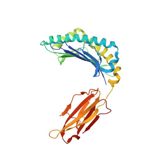
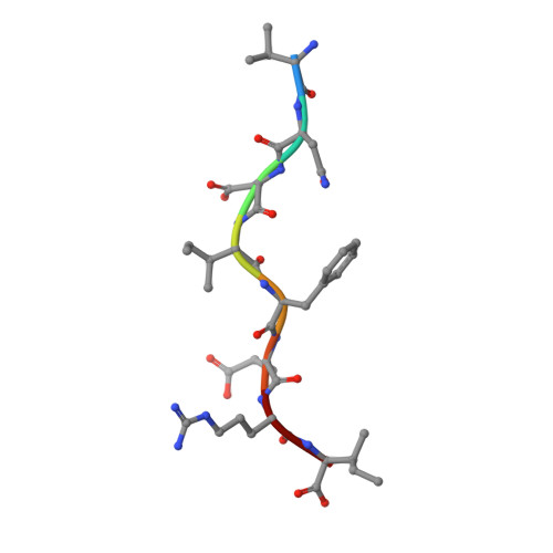
Crystal structure of the Class I MHC Molecule H-2Kwm7 with a Single Self Peptide VNDIFERI


Experimental Data Snapshot

  • Method: X-RAY DIFFRACTION
  • Resolution: 2.50 Å
  • R-Value Free: 
    0.261 (Depositor), 0.248 (DCC) 
  • R-Value Work: 
    0.192 (Depositor), 0.197 (DCC) 
  • R-Value Observed: 
    0.196 (Depositor) 

wwPDB Validation 3D Report Full Report

Validation slider image for 3FOL

This is version 1.3 of the entry. See complete history. 

Literature

Predominant occupation of the class I MHC molecule H-2Kwm7 with a single self-peptide suggests a mechanism for its diabetes-protective effect

Brims, D.R., Qian, J., Jarchum, I., Mikesh, L., Palmieri, E., Ramagopal, U.A., Malashkevich, V.N., Chaparro, R.J., Lund, T., Hattori, M., Shabanowitz, J., Hunt, D.F., Nathenson, S.G., Almo, S.C., Dilorenzo, T.P.

(2010) Int Immunol 22: 191-203

  • DOI: https://doi.org/10.1093/intimm/dxp127
  • Primary Citation Related Structures: 
    3FOL, 3FOM, 3FON

  • PubMed Abstract: 

    Type 1 diabetes (T1D) is an autoimmune disease characterized by T cell-mediated destruction of insulin-producing pancreatic beta cells. In both humans and the non-obese diabetic (NOD) mouse model of T1D, class II MHC alleles are the primary determinant of disease susceptibility. However, class I MHC genes also influence risk. These findings are consistent with the requirement for both CD4(+) and CD8(+) T cells in the pathogenesis of T1D. Although a large body of work has permitted the identification of multiple mechanisms to explain the diabetes-protective effect of particular class II MHC alleles, studies examining the protective influence of class I alleles are lacking. Here, we explored this question by performing biochemical and structural analyses of the murine class I MHC molecule H-2K(wm7), which exerts a diabetes-protective effect in NOD mice. We have found that H-2K(wm7) molecules are predominantly occupied by the single self-peptide VNDIFERI, derived from the ubiquitous protein histone H2B. This unexpected finding suggests that the inability of H-2K(wm7) to support T1D development could be due, at least in part, to the failure of peptides from critical beta-cell antigens to adequately compete for binding and be presented to T cells. Predominant presentation of a single peptide would also be expected to influence T-cell selection, potentially leading to a reduced ability to select a diabetogenic CD8(+) T-cell repertoire. The report that one of the predominant peptides bound by T1D-protective HLA-A*31 is histone derived suggests the potential translation of our findings to human diabetes-protective class I MHC molecules.


  • Organizational Affiliation: 
    • Department of Microbiology and Immunology, Albert Einstein College of Medicine, Bronx, NY 10461, USA.

Macromolecule Content 

  • Total Structure Weight: 44.33 kDa 
  • Atom Count: 3,196 
  • Modeled Residue Count: 382 
  • Deposited Residue Count: 382 
  • Unique protein chains: 3

Macromolecules

Find similar proteins by:|  3D Structure
Entity ID: 1
MoleculeChains  Sequence LengthOrganismDetailsImage
MHC274Mus musculusMutation(s): 0 
UniProt
Find proteins for D2YW38 (Mus musculus)
Explore D2YW38 
Go to UniProtKB:  D2YW38
Entity Groups
Sequence Clusters30% Identity50% Identity70% Identity90% Identity95% Identity100% Identity
UniProt GroupD2YW38
Sequence Annotations
Expand
Reference Sequence
Find similar proteins by:|  3D Structure
Entity ID: 2
MoleculeChains  Sequence LengthOrganismDetailsImage
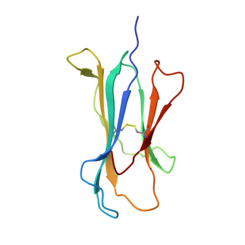
Beta-2-microglobulin100Mus musculusMutation(s): 0 
Gene Names: B2m
UniProt & NIH Common Fund Data Resources
Find proteins for P01887 (Mus musculus)
Explore P01887 
Go to UniProtKB:  P01887
IMPC:  MGI:88127
Entity Groups
Sequence Clusters30% Identity50% Identity70% Identity90% Identity95% Identity100% Identity
UniProt GroupP01887
Sequence Annotations
Expand
Reference Sequence
Find similar proteins by:  Sequence   |   3D Structure  
Entity ID: 3
MoleculeChains  Sequence LengthOrganismDetailsImage
8 residue synthetic peptideC [auth P]8N/AMutation(s): 0 
Sequence Annotations
Expand
Reference Sequence

Experimental Data & Validation

Experimental Data

  • Method: X-RAY DIFFRACTION
  • Resolution: 2.50 Å
  • R-Value Free:  0.261 (Depositor), 0.248 (DCC) 
  • R-Value Work:  0.192 (Depositor), 0.197 (DCC) 
  • R-Value Observed: 0.196 (Depositor) 
Space Group: C 1 2 1
Unit Cell:
Length ( Å )Angle ( ˚ )
a = 117.393α = 90
b = 66.63β = 93.66
c = 56.211γ = 90
Software Package:
Software NamePurpose
DENZOdata reduction
SCALEPACKdata scaling
REFMACrefinement
PDB_EXTRACTdata extraction
StructureStudiodata collection
HKL-2000data reduction
PHASERphasing

Structure Validation

View Full Validation Report


Entry History 

Revision History  (Full details and data files)

  • Version 1.0: 2010-01-12
    Type: Initial release
  • Version 1.1: 2011-07-13
    Changes: Advisory, Refinement description, Version format compliance
  • Version 1.2: 2017-11-01
    Changes: Refinement description
  • Version 1.3: 2024-11-06
    Changes: Data collection, Database references, Structure summary