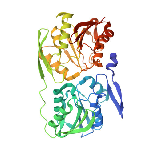
Crystal Structure of the Thermus thermophilus 16 S rRNA Methyltransferase RsmC in Complex with Cofactor and Substrate Guanosine.
Demirci, H., Gregory, S.T., Dahlberg, A.E., Jogl, G.(2008) J Biol Chem 283: 26548-26556
- PubMed: 18667428 Search on PubMedSearch on PubMed Central
- DOI: https://doi.org/10.1074/jbc.M804005200
- Primary Citation Related Structures:
3DMF, 3DMG, 3DMH - PubMed Abstract:
Post-transcriptional modification is a ubiquitous feature of ribosomal RNA in all kingdoms of life. Modified nucleotides are generally clustered in functionally important regions of the ribosome, but the functional contribution to protein synthesis is not well understood. Here we describe high resolution crystal structures for the N(2)-guanine methyltransferase RsmC that modifies residue G1207 in 16 S rRNA near the decoding site of the 30 S ribosomal subunit. RsmC is a class I S-adenosyl-L-methionine-dependent methyltransferase composed of two methyltransferase domains. However, only one S-adenosyl-L-methionine molecule and one substrate molecule, guanosine, bind in the ternary complex. The N-terminal domain does not bind any cofactor. Two structures with bound S-adenosyl-L-methionine and S-adenosyl-L-homocysteine confirm that the cofactor binding mode is highly similar to other class I methyltransferases. Secondary structure elements of the N-terminal domain contribute to cofactor-binding interactions and restrict access to the cofactor-binding site. The orientation of guanosine in the active site reveals that G1207 has to disengage from its Watson-Crick base pairing interaction with C1051 in the 16 S rRNA and flip out into the active site prior to its modification. Inspection of the 30 S crystal structure indicates that access to G1207 by RsmC is incompatible with the native subunit structure, consistent with previous suggestions that this enzyme recognizes a subunit assembly intermediate.
- Department of Molecular Biology, Cell Biology and Biochemistry, Brown University, Providence, Rhode Island 02912, USA.
Organizational Affiliation: