3D3T | pdb_00003d3t

Crystal Structure of HIV-1 CRF01_AE in complex with the substrate p1-p6


Experimental Data Snapshot

  • Method: X-RAY DIFFRACTION
  • Resolution: 2.80 Å
  • R-Value Free: 
    0.258 (Depositor), 0.221 (DCC) 
  • R-Value Work: 
    0.199 (Depositor), 0.194 (DCC) 
  • R-Value Observed: 
    0.203 (Depositor) 

wwPDB Validation 3D Report Full Report

Validation slider image for 3D3T

This is version 1.4 of the entry. See complete history

Literature

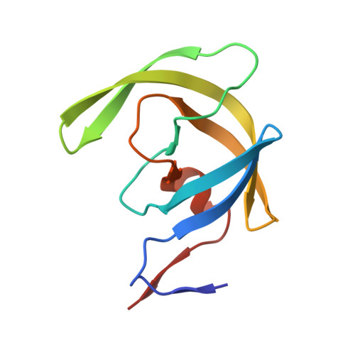
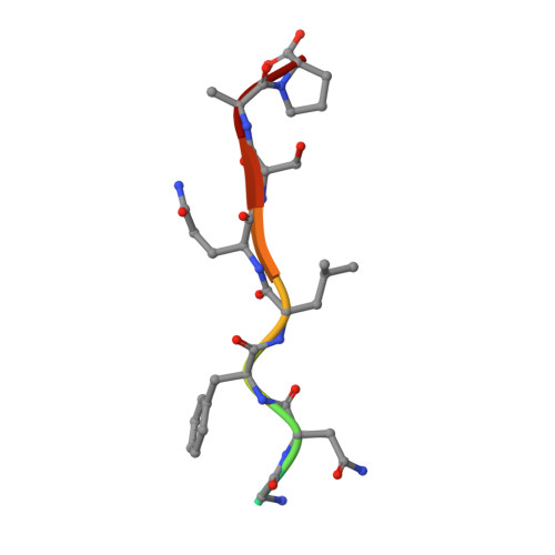
Structural analysis of human immunodeficiency virus type 1 CRF01_AE protease in complex with the substrate p1-p6.

Bandaranayake, R.M.Prabu-Jeyabalan, M.Kakizawa, J.Sugiura, W.Schiffer, C.A.

(2008) J Virol 82: 6762-6766

  • DOI: https://doi.org/10.1128/JVI.00018-08
  • Primary Citation Related Structures: 
    3D3T

  • PubMed Abstract: 

    The effect of amino acid variability between human immunodeficiency virus type 1 (HIV-1) clades on structure and the emergence of resistance mutations in HIV-1 protease has become an area of significant interest in recent years. We determined the first crystal structure of the HIV-1 CRF01_AE protease in complex with the p1-p6 substrate to a resolution of 2.8 A. Hydrogen bonding between the flap hinge and the protease core regions shows significant structural rearrangements in CRF01_AE protease compared to the clade B protease structure.


  • Organizational Affiliation
    • Department of Biochemistry and Molecular Pharmacology, University of Massachusetts Medical School, 364 Plantation Street, Worcester, MA 01605, USA.

Macromolecule Content 

  • Total Structure Weight: 22.79 kDa 
  • Atom Count: 1,494 
  • Modeled Residue Count: 206 
  • Deposited Residue Count: 208 
  • Unique protein chains: 2

Macromolecules

Find similar proteins by:|  3D Structure
Entity ID: 1
MoleculeChains  Sequence LengthOrganismDetailsImage
HIV-1 protease
A, B
99Human immunodeficiency virus 1Mutation(s): 1 
Gene Names: gag-pol
EC: 3.4.23.16
UniProt
Find proteins for Q90VT5 (Human immunodeficiency virus type 1)
Explore Q90VT5 
Go to UniProtKB:  Q90VT5
Entity Groups
Sequence Clusters30% Identity50% Identity70% Identity90% Identity95% Identity100% Identity
UniProt GroupQ90VT5
Sequence Annotations
Expand
Reference Sequence
Find similar proteins by:  Sequence   |   3D Structure  
Entity ID: 2
MoleculeChains  Sequence LengthOrganismDetailsImage
P1-P6 SUBSTRATE PEPTIDEC [auth P]10N/AMutation(s): 0 
UniProt
Find proteins for P12495 (Human immunodeficiency virus type 1 group M subtype D (isolate Z2/CDC-Z34))
Explore P12495 
Go to UniProtKB:  P12495
Entity Groups
Sequence Clusters30% Identity50% Identity70% Identity90% Identity95% Identity100% Identity
UniProt GroupP12495
Sequence Annotations
Expand
Reference Sequence

Experimental Data & Validation

Experimental Data

  • Method: X-RAY DIFFRACTION
  • Resolution: 2.80 Å
  • R-Value Free:  0.258 (Depositor), 0.221 (DCC) 
  • R-Value Work:  0.199 (Depositor), 0.194 (DCC) 
  • R-Value Observed: 0.203 (Depositor) 
Space Group: P 61
Unit Cell:
Length ( Å )Angle ( ˚ )
a = 62.186α = 90
b = 62.186β = 90
c = 82.099γ = 120
Software Package:
Software NamePurpose
DENZOdata reduction
SCALEPACKdata scaling
AMoREphasing
REFMACrefinement
PDB_EXTRACTdata extraction

Structure Validation

View Full Validation Report


Entry History 

Deposition Data

Revision History  (Full details and data files)

  • Version 1.0: 2008-07-08
    Type: Initial release
  • Version 1.1: 2011-07-13
    Changes: Advisory, Version format compliance
  • Version 1.2: 2017-10-25
    Changes: Refinement description
  • Version 1.3: 2021-10-20
    Changes: Database references
  • Version 1.4: 2024-02-21
    Changes: Data collection