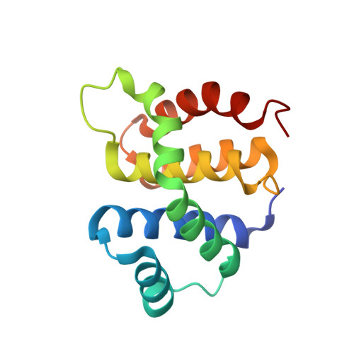
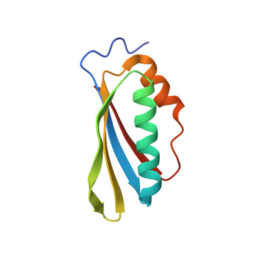
3D3B

Structural and functional analysis of the E. coli NusB-S10 transcription antitermination complex.


Experimental Data Snapshot

  • Method: X-RAY DIFFRACTION
  • Resolution: 1.30 Å
  • R-Value Free: 0.204 
  • R-Value Work: 0.173 
  • R-Value Observed: 0.175 

Starting Models: experimental
View more details

wwPDB Validation   3D Report Full Report


This is version 1.4 of the entry. See complete history


Literature

Structural and functional analysis of the E. coli NusB-S10 transcription antitermination complex.

Luo, X.Hsiao, H.H.Bubunenko, M.Weber, G.Court, D.L.Gottesman, M.E.Urlaub, H.Wahl, M.C.

(2008) Mol Cell 32: 791-802

  • DOI: https://doi.org/10.1016/j.molcel.2008.10.028
  • Primary Citation of Related Structures:  
    3D3B, 3D3C

  • PubMed Abstract: 

    Protein S10 is a component of the 30S ribosomal subunit and participates together with NusB protein in processive transcription antitermination. The molecular mechanisms by which S10 can act as a translation or a transcription factor are not understood. We used complementation assays and recombineering to delineate regions of S10 dispensable for antitermination, and determined the crystal structure of a transcriptionally active NusB-S10 complex. In this complex, S10 adopts the same fold as in the 30S subunit and is blocked from simultaneous association with the ribosome. Mass spectrometric mapping of UV-induced crosslinks revealed that the NusB-S10 complex presents an intermolecular, composite, and contiguous binding surface for RNAs containing BoxA antitermination signals. Furthermore, S10 overproduction complemented a nusB null phenotype. These data demonstrate that S10 and NusB together form a BoxA-binding module, that NusB facilitates entry of S10 into the transcription machinery, and that S10 represents a central hub in processive antitermination.


  • Organizational Affiliation

    Research Group X-Ray Crystallography, Max-Planck-Institute for Biophysical Chemistry, D-37077 Göttingen, Germany.


Macromolecules
Find similar proteins by:  (by identity cutoff)  |  3D Structure
Entity ID: 1
MoleculeChains Sequence LengthOrganismDetailsImage
N utilization substance protein B141Escherichia coli K-12Mutation(s): 1 
Gene Names: nusBssyBb0416JW0406
UniProt
Find proteins for P0A780 (Escherichia coli (strain K12))
Explore P0A780 
Go to UniProtKB:  P0A780
Entity Groups  
Sequence Clusters30% Identity50% Identity70% Identity90% Identity95% Identity100% Identity
UniProt GroupP0A780
Sequence Annotations
Expand
  • Reference Sequence
Find similar proteins by:  (by identity cutoff)  |  3D Structure
Entity ID: 2
MoleculeChains Sequence LengthOrganismDetailsImage
30S ribosomal protein S10B [auth J]87Escherichia coli K-12Mutation(s): 0 
Gene Names: rpsJnusEb3321JW3283
UniProt
Find proteins for P0A7R5 (Escherichia coli (strain K12))
Explore P0A7R5 
Go to UniProtKB:  P0A7R5
Entity Groups  
Sequence Clusters30% Identity50% Identity70% Identity90% Identity95% Identity100% Identity
UniProt GroupP0A7R5
Sequence Annotations
Expand
  • Reference Sequence
Experimental Data & Validation

Experimental Data

  • Method: X-RAY DIFFRACTION
  • Resolution: 1.30 Å
  • R-Value Free: 0.204 
  • R-Value Work: 0.173 
  • R-Value Observed: 0.175 
  • Space Group: P 21 21 21
Unit Cell:
Length ( Å )Angle ( ˚ )
a = 40.74α = 90
b = 48.99β = 90
c = 112.77γ = 90
Software Package:
Software NamePurpose
REFMACrefinement
MAR345dtbdata collection
XDSdata reduction
XDSdata scaling
MOLREPphasing

Structure Validation

View Full Validation Report


Entry History 

Deposition Data

Revision History  (Full details and data files)

  • Version 1.0: 2009-01-13
    Type: Initial release
  • Version 1.1: 2011-07-13
    Changes: Advisory, Source and taxonomy, Version format compliance
  • Version 1.2: 2017-08-02
    Changes: Source and taxonomy
  • Version 1.3: 2021-10-20
    Changes: Database references, Derived calculations
  • Version 1.4: 2023-08-30
    Changes: Data collection, Refinement description