3D23

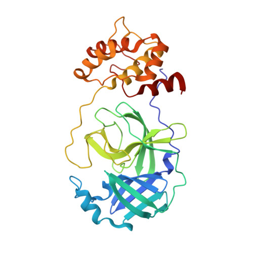
Main protease of HCoV-HKU1


Experimental Data Snapshot

  • Method: X-RAY DIFFRACTION
  • Resolution: 2.50 Å
  • R-Value Free: 0.285 
  • R-Value Work: 0.229 
  • R-Value Observed: 0.232 

wwPDB Validation   3D Report Full Report


This is version 2.1 of the entry. See complete history


Literature

Structure of the main protease from a global infectious human coronavirus, HCoV-HKU1.

Zhao, Q.Li, S.Xue, F.Zou, Y.Chen, C.Bartlam, M.Rao, Z.

(2008) J Virol 82: 8647-8655

  • DOI: https://doi.org/10.1128/JVI.00298-08
  • Primary Citation of Related Structures:  
    3D23

  • PubMed Abstract: 

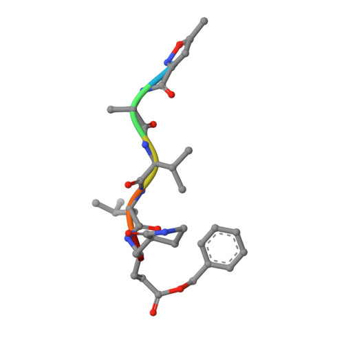
    The newly emergent human coronavirus HKU1 (HCoV-HKU1) was first identified in Hong Kong in 2005. Infection by HCoV-HKU1 occurs worldwide and causes syndromes such as the common cold, bronchitis, and pneumonia. The CoV main protease (M(pro)), which is a key enzyme in viral replication via the proteolytic processing of the replicase polyproteins, has been recognized as an attractive target for rational drug design. In this study, we report the structure of HCoV-HKU1 M(pro) in complex with a Michael acceptor, inhibitor N3. The structure of HCoV-HKU1 provides a high-quality model for group 2A CoVs, which are distinct from group 2B CoVs such as severe acute respiratory syndrome CoV. The structure, together with activity assays, supports the relative conservation at the P1 position that was discovered by sequencing the HCoV-HKU1 genome. Combined with structural data from other CoV M(pro)s, the HCoV-HKU1 M(pro) structure reported here provides insights into both substrate preference and the design of antivirals targeting CoVs.


  • Organizational Affiliation

    Tsinghua-Nankai-IBP Joint Research Group for Structural Biology, Tsinghua University, Beijing, China.


Macromolecules
Find similar proteins by:  (by identity cutoff)  |  3D Structure
Entity ID: 1
MoleculeChains Sequence LengthOrganismDetailsImage
3C-like proteinaseA [auth B],
B [auth A],
C,
D
302Human coronavirus HKU1 (isolate N1)Mutation(s): 0 
Gene Names: rep
EC: 3.4.22
UniProt
Find proteins for P0C6U3 (Human coronavirus HKU1 (isolate N1))
Explore P0C6U3 
Go to UniProtKB:  P0C6U3
Entity Groups  
Sequence Clusters30% Identity50% Identity70% Identity90% Identity95% Identity100% Identity
UniProt GroupP0C6U3
Sequence Annotations
Expand
  • Reference Sequence

Find similar proteins by:  Sequence   |   3D Structure  

Entity ID: 2
MoleculeChains Sequence LengthOrganismDetailsImage
N-[(5-METHYLISOXAZOL-3-YL)CARBONYL]ALANYL-L-VALYL-N~1~-((1R,2Z)-4-(BENZYLOXY)-4-OXO-1-{[(3R)-2-OXOPYRROLIDIN-3-YL]METHYL}BUT-2-ENYL)-L-LEUCINAMIDEE [auth H],
F,
G [auth E],
H [auth G]
6synthetic constructMutation(s): 0 
Sequence Annotations
Expand
  • Reference Sequence
Experimental Data & Validation

Experimental Data

  • Method: X-RAY DIFFRACTION
  • Resolution: 2.50 Å
  • R-Value Free: 0.285 
  • R-Value Work: 0.229 
  • R-Value Observed: 0.232 
  • Space Group: P 41
Unit Cell:
Length ( Å )Angle ( ˚ )
a = 91.77α = 90
b = 91.77β = 90
c = 187.914γ = 90
Software Package:
Software NamePurpose
REFMACrefinement
PDB_EXTRACTdata extraction
DENZOdata reduction
SCALEPACKdata scaling

Structure Validation

View Full Validation Report


Entry History 

Deposition Data

Revision History  (Full details and data files)

  • Version 1.0: 2008-09-16
    Type: Initial release
  • Version 1.1: 2011-07-13
    Changes: Source and taxonomy, Version format compliance
  • Version 1.2: 2015-12-09
    Changes: Derived calculations, Structure summary
  • Version 1.3: 2017-10-25
    Changes: Refinement description
  • Version 2.0: 2023-11-15
    Changes: Atomic model, Data collection, Database references, Derived calculations
  • Version 2.1: 2024-10-30
    Changes: Structure summary