3CQA | pdb_00003cqa

Crystal structure of human fibroblast growth factor-1 with mutations Glu81Ala and Lys101Ala


Experimental Data Snapshot

  • Method: X-RAY DIFFRACTION
  • Resolution: 1.80 Å
  • R-Value Free: 
    0.212 (Depositor), 0.221 (DCC) 
  • R-Value Work: 
    0.191 (Depositor), 0.198 (DCC) 
  • R-Value Observed: 
    0.191 (Depositor) 

Starting Model: experimental
View more details

wwPDB Validation 3D Report Full Report

Validation slider image for 3CQA

This is version 1.3 of the entry. See complete history

Literature

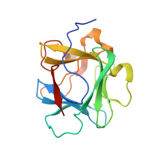
Engineering an improved crystal contact across a solvent-mediated interface of human fibroblast growth factor 1.

Meher, A.K.Blaber, S.I.Lee, J.Honjo, E.Kuroki, R.Blaber, M.

(2009) Acta Crystallogr Sect F Struct Biol Cryst Commun 65: 1136-1140

  • DOI: https://doi.org/10.1107/S1744309109036987
  • Primary Citation Related Structures: 
    3CQA, 3CRG, 3CRH, 3CRI

  • PubMed Abstract: 

    Large-volume protein crystals are a prerequisite for neutron diffraction studies and their production represents a bottleneck in obtaining neutron structures. Many protein crystals that permit the collection of high-resolution X-ray diffraction data are inappropriate for neutron diffraction owing to a plate-type morphology that limits the crystal volume. Human fibroblast growth factor 1 crystallizes in a plate morphology that yields atomic resolution X-ray diffraction data but has insufficient volume for neutron diffraction. The thin physical dimension has been identified as corresponding to the b cell edge and the X-ray structure identified a solvent-mediated crystal contact adjacent to position Glu81 that was hypothesized to limit efficient crystal growth in this dimension. In this report, a series of mutations at this crystal contact designed to both reduce side-chain entropy and replace the solvent-mediated interface with direct side-chain contacts are reported. The results suggest that improved crystal growth is achieved upon the introduction of direct crystal contacts, while little improvement is observed with side-chain entropy-reducing mutations alone.


  • Organizational Affiliation
    • Department of Biomedical Sciences, Florida State University, Tallahassee, 32306-4300, USA.

Macromolecule Content 

  • Total Structure Weight: 32.87 kDa 
  • Atom Count: 2,482 
  • Modeled Residue Count: 281 
  • Deposited Residue Count: 288 
  • Unique protein chains: 1

Macromolecules

Find similar proteins by:|  3D Structure
Entity ID: 1
MoleculeChains  Sequence LengthOrganismDetailsImage
Heparin-binding growth factor 1
A, B
144Homo sapiensMutation(s): 2 
Gene Names: FGF1FGFA
UniProt & NIH Common Fund Data Resources
Find proteins for P05230 (Homo sapiens)
Explore P05230 
Go to UniProtKB:  P05230
PHAROS:  P05230
GTEx:  ENSG00000113578 
Entity Groups
Sequence Clusters30% Identity50% Identity70% Identity90% Identity95% Identity100% Identity
UniProt GroupP05230
Sequence Annotations
Expand
Reference Sequence

Experimental Data & Validation

Experimental Data

  • Method: X-RAY DIFFRACTION
  • Resolution: 1.80 Å
  • R-Value Free:  0.212 (Depositor), 0.221 (DCC) 
  • R-Value Work:  0.191 (Depositor), 0.198 (DCC) 
  • R-Value Observed: 0.191 (Depositor) 
Space Group: C 2 2 21
Unit Cell:
Length ( Å )Angle ( ˚ )
a = 73.34α = 90
b = 97.91β = 90
c = 108.48γ = 90
Software Package:
Software NamePurpose
CNSrefinement
HKL-2000data collection
HKL-2000data reduction
HKL-2000data scaling
CNSphasing

Structure Validation

View Full Validation Report


Entry History 

Deposition Data

Revision History  (Full details and data files)

  • Version 1.0: 2009-04-07
    Type: Initial release
  • Version 1.1: 2011-07-13
    Changes: Version format compliance
  • Version 1.2: 2021-10-20
    Changes: Database references, Derived calculations
  • Version 1.3: 2023-08-30
    Changes: Data collection, Refinement description