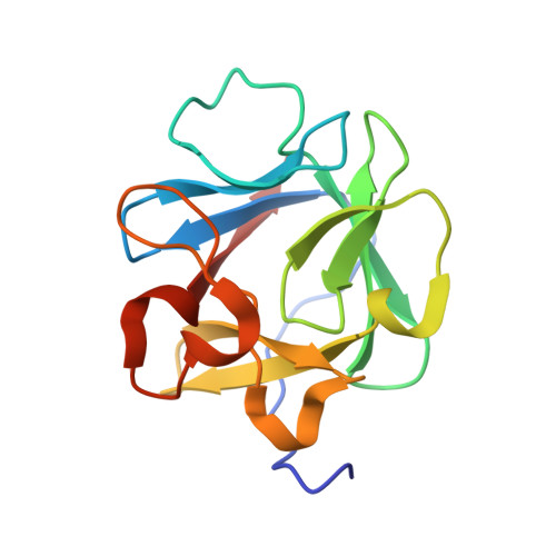
Engineering an improved crystal contact across a solvent-mediated interface of human fibroblast growth factor 1.
Meher, A.K., Blaber, S.I., Lee, J., Honjo, E., Kuroki, R., Blaber, M.(2009) Acta Crystallogr Sect F Struct Biol Cryst Commun 65: 1136-1140
- PubMed: 19923735
- DOI: https://doi.org/10.1107/S1744309109036987
- Primary Citation of Related Structures:
3CQA, 3CRG, 3CRH, 3CRI - PubMed Abstract:
Large-volume protein crystals are a prerequisite for neutron diffraction studies and their production represents a bottleneck in obtaining neutron structures. Many protein crystals that permit the collection of high-resolution X-ray diffraction data are inappropriate for neutron diffraction owing to a plate-type morphology that limits the crystal volume. Human fibroblast growth factor 1 crystallizes in a plate morphology that yields atomic resolution X-ray diffraction data but has insufficient volume for neutron diffraction. The thin physical dimension has been identified as corresponding to the b cell edge and the X-ray structure identified a solvent-mediated crystal contact adjacent to position Glu81 that was hypothesized to limit efficient crystal growth in this dimension. In this report, a series of mutations at this crystal contact designed to both reduce side-chain entropy and replace the solvent-mediated interface with direct side-chain contacts are reported. The results suggest that improved crystal growth is achieved upon the introduction of direct crystal contacts, while little improvement is observed with side-chain entropy-reducing mutations alone.
Organizational Affiliation:
Department of Biomedical Sciences, Florida State University, Tallahassee, 32306-4300, USA.


