3BUS | pdb_00003bus

Crystal Structure of RebM


Experimental Data Snapshot

  • Method: X-RAY DIFFRACTION
  • Resolution: 2.65 Å
  • R-Value Free: 
    0.260 (Depositor) 
  • R-Value Work: 
    0.214 (Depositor), 0.225 (DCC) 
  • R-Value Observed: 
    0.217 (Depositor) 

wwPDB Validation 3D Report Full Report

Validation slider image for 3BUS

This is version 1.4 of the entry. See complete history

Literature

Structure and mechanism of the rebeccamycin sugar 4'-O-methyltransferase RebM.

Singh, S.McCoy, J.G.Zhang, C.Bingman, C.A.Phillips Jr., G.N.Thorson, J.S.

(2008) J Biological Chem 283: 22628-22636

  • DOI: https://doi.org/10.1074/jbc.M800503200
  • Primary Citation Related Structures: 
    3BUS

  • PubMed Abstract: 

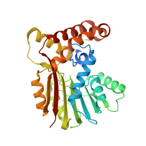
    The 2.65-angstroms crystal structure of the rebeccamycin 4'-O-methyltransferase RebM in complex with S-adenosyl-l-homocysteine revealed RebM to adopt a typical S-adenosylmethionine-binding fold of small molecule O-methyltransferases (O-MTases) and display a weak dimerization domain unique to MTases. Using this structure as a basis, the RebM substrate binding model implicated a predominance of nonspecific hydrophobic interactions consistent with the reported ability of RebM to methylate a wide range of indolocarbazole surrogates. This model also illuminated the three putative RebM catalytic residues (His140/141 and Asp166) subsequently found to be highly conserved among sequence-related natural product O-MTases from GC-rich bacteria. Interrogation of these residues via site-directed mutagenesis in RebM demonstrated His140 and Asp166 to be most important for catalysis. This study reveals RebM to be a member of the general acid/base-dependent O-MTases and, as the first crystal structure for a sugar O-MTase, may also present a template toward the future engineering of natural product MTases for combinatorial applications.


  • Organizational Affiliation
    • Division of Pharmaceutical Sciences, School of Pharmacy, University of Wisconsin, Madison, Wisconsin 53705, USA.

Macromolecule Content 

  • Total Structure Weight: 60.35 kDa 
  • Atom Count: 3,767 
  • Modeled Residue Count: 493 
  • Deposited Residue Count: 546 
  • Unique protein chains: 1

Macromolecules

Find similar proteins by:|  3D Structure
Entity ID: 1
MoleculeChains  Sequence LengthOrganismDetailsImage
Methyltransferase
A, B
273Lentzea aerocolonigenesMutation(s): 0 
Gene Names: rbmFrebM
EC: 2.1.1.164
UniProt
Find proteins for Q8KZ94 (Lentzea aerocolonigenes)
Explore Q8KZ94 
Go to UniProtKB:  Q8KZ94
Entity Groups
Sequence Clusters30% Identity50% Identity70% Identity90% Identity95% Identity100% Identity
UniProt GroupQ8KZ94
Sequence Annotations
Expand
Reference Sequence

Small Molecules

Ligands 1 Unique
IDChains Name / Formula / InChI Key2D Diagram3D Interactions
SAH

Query on SAH


Download:Ideal Coordinates CCD File
C [auth A],
D [auth B]
S-ADENOSYL-L-HOMOCYSTEINE
C14 H20 N6 O5 S
ZJUKTBDSGOFHSH-WFMPWKQPSA-N
Modified Residues  1 Unique
IDChains TypeFormula2D DiagramParent
MSE
Query on MSE
A, B
L-PEPTIDE LINKINGC5 H11 N O2 SeMET

Experimental Data & Validation

Experimental Data

  • Method: X-RAY DIFFRACTION
  • Resolution: 2.65 Å
  • R-Value Free:  0.260 (Depositor) 
  • R-Value Work:  0.214 (Depositor), 0.225 (DCC) 
  • R-Value Observed: 0.217 (Depositor) 
Space Group: P 41 21 2
Unit Cell:
Length ( Å )Angle ( ˚ )
a = 119.153α = 90
b = 119.153β = 90
c = 84.412γ = 90
Software Package:
Software NamePurpose
DENZOdata reduction
SCALEPACKdata scaling
SHARPphasing
DMphasing
REFMACrefinement
PDB_EXTRACTdata extraction

Structure Validation

View Full Validation Report


Entry History 

Deposition Data

Revision History  (Full details and data files)

  • Version 1.0: 2008-03-25
    Type: Initial release
  • Version 1.1: 2011-07-13
    Changes: Advisory, Version format compliance
  • Version 1.2: 2013-01-30
    Changes: Database references, Structure summary
  • Version 1.3: 2017-10-25
    Changes: Refinement description
  • Version 1.4: 2024-10-30
    Changes: Data collection, Database references, Derived calculations, Structure summary