3B95

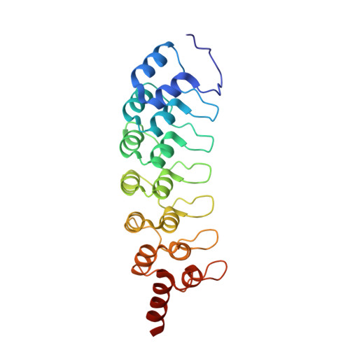
EuHMT1 (Glp) Ankyrin Repeat Domain (Structure 2)


Experimental Data Snapshot

  • Method: X-RAY DIFFRACTION
  • Resolution: 2.99 Å
  • R-Value Free: 0.238 
  • R-Value Work: 0.195 
  • R-Value Observed: 0.198 

Starting Model: experimental
View more details

wwPDB Validation   3D Report Full Report


This is version 1.3 of the entry. See complete history


Literature

The ankyrin repeats of G9a and GLP histone methyltransferases are mono- and dimethyllysine binding modules.

Collins, R.E.Northrop, J.P.Horton, J.R.Lee, D.Y.Zhang, X.Stallcup, M.R.Cheng, X.

(2008) Nat Struct Mol Biol 15: 245-250

  • DOI: https://doi.org/10.1038/nsmb.1384
  • Primary Citation of Related Structures:  
    3B7B, 3B95

  • PubMed Abstract: 

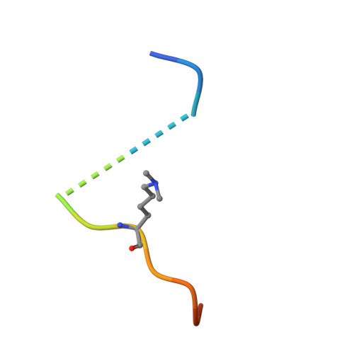
    Histone modifications have important roles in transcriptional control, mitosis and heterochromatin formation. G9a and G9a-like protein (GLP) are euchromatin-associated methyltransferases that repress transcription by mono- and dimethylating histone H3 at Lys9 (H3K9). Here we demonstrate that the ankyrin repeat domains of G9a and GLP bind with strong preference to N-terminal H3 peptides containing mono- or dimethyl K9. X-ray crystallography revealed the basis for recognition of the methylated lysine by a partial hydrophobic cage with three tryptophans and one acidic residue. Substitution of key residues in the cage eliminated the H3 tail interaction. Hence, G9a and GLP contain a new type of methyllysine binding module (the ankyrin repeat domains) and are the first examples of protein (histone) methyltransferases harboring in a single polypeptide the activities that generate and read the same epigenetic mark.


  • Organizational Affiliation

    Department of Biochemistry, Emory University School of Medicine, 1510 Clifton Road, Atlanta, Georgia 30322, USA.


Macromolecules
Find similar proteins by:  (by identity cutoff)  |  3D Structure
Entity ID: 1
MoleculeChains Sequence LengthOrganismDetailsImage
Euchromatic histone-lysine N-methyltransferase 1
A, B
237Homo sapiensMutation(s): 0 
Gene Names: EHMT1
EC: 2.1.1 (UniProt), 2.1.1.367 (UniProt)
UniProt & NIH Common Fund Data Resources
Find proteins for Q9H9B1 (Homo sapiens)
Explore Q9H9B1 
Go to UniProtKB:  Q9H9B1
PHAROS:  Q9H9B1
GTEx:  ENSG00000181090 
Entity Groups  
Sequence Clusters30% Identity50% Identity70% Identity90% Identity95% Identity100% Identity
UniProt GroupQ9H9B1
Sequence Annotations
Expand
  • Reference Sequence

Find similar proteins by:  Sequence   |   3D Structure  

Entity ID: 2
MoleculeChains Sequence LengthOrganismDetailsImage
Histone H3 N-terminal PeptideC [auth P]15Homo sapiensMutation(s): 0 
Gene Names: H3C1H3C2H3C3H3C4H3C6H3C7H3C8H3C10H3C11H3C12
UniProt & NIH Common Fund Data Resources
Find proteins for P68431 (Homo sapiens)
Explore P68431 
Go to UniProtKB:  P68431
PHAROS:  P68431
Entity Groups  
Sequence Clusters30% Identity50% Identity70% Identity90% Identity95% Identity100% Identity
UniProt GroupP68431
Sequence Annotations
Expand
  • Reference Sequence
Small Molecules
Modified Residues  1 Unique
IDChains TypeFormula2D DiagramParent
MLY
Query on MLY
C [auth P]L-PEPTIDE LINKINGC8 H18 N2 O2LYS
Experimental Data & Validation

Experimental Data

  • Method: X-RAY DIFFRACTION
  • Resolution: 2.99 Å
  • R-Value Free: 0.238 
  • R-Value Work: 0.195 
  • R-Value Observed: 0.198 
  • Space Group: C 2 2 21
Unit Cell:
Length ( Å )Angle ( ˚ )
a = 59.52α = 90
b = 152.39β = 90
c = 167.32γ = 90
Software Package:
Software NamePurpose
MAR345dtbdata collection
GLRFphasing
CNSrefinement
HKL-2000data reduction
HKL-2000data scaling

Structure Validation

View Full Validation Report


Entry History 

Deposition Data

Revision History  (Full details and data files)

  • Version 1.0: 2008-02-12
    Type: Initial release
  • Version 1.1: 2011-07-13
    Changes: Version format compliance
  • Version 1.2: 2012-02-01
    Changes: Derived calculations
  • Version 1.3: 2023-08-30
    Changes: Data collection, Database references, Derived calculations, Refinement description