3ADC

Crystal structure of O-phosphoseryl-tRNA kinase complexed with selenocysteine tRNA and AMPPNP (crystal type 2)


Experimental Data Snapshot

  • Method: X-RAY DIFFRACTION
  • Resolution: 2.90 Å
  • R-Value Free: 0.277 
  • R-Value Work: 0.224 
  • R-Value Observed: 0.227 

Starting Model: experimental
View more details

wwPDB Validation   3D Report Full Report


Ligand Structure Quality Assessment 


This is version 1.3 of the entry. See complete history


Literature

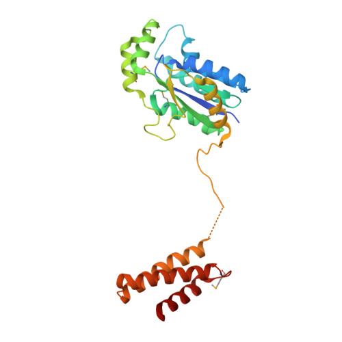
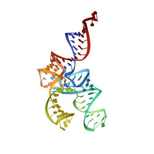
Structural Basis for the Major Role of O-Phosphoseryl-tRNA Kinase in the UGA-Specific Encoding of Selenocysteine

Chiba, S.Itoh, Y.Sekine, S.Yokoyama, S.

(2010) Mol Cell 39: 410-420

  • DOI: https://doi.org/10.1016/j.molcel.2010.07.018
  • Primary Citation of Related Structures:  
    3ADB, 3ADC, 3ADD

  • PubMed Abstract: 

    The 21(st) amino acid, selenocysteine (Sec), is assigned to the codon UGA and is biosynthesized on the selenocysteine-specific tRNA (tRNA(Sec)) with the corresponding anticodon. In archaea/eukarya, tRNA(Sec) is ligated with serine by seryl-tRNA synthetase (SerRS), the seryl moiety is phosphorylated by O-phosphoseryl-tRNA kinase (PSTK), and the phosphate group is replaced with selenol by Sep-tRNA:Sec-tRNA synthase. PSTK selectively phosphorylates seryl-tRNA(Sec), while SerRS serylates both tRNA(Ser) and tRNA(Sec). In this study, we determined the crystal structures of the archaeal tRNA(Sec).PSTK complex. PSTK consists of two independent linker-connected domains, the N-terminal catalytic domain (NTD) and the C-terminal domain (CTD). The D-arm.CTD binding occurs independently of and much more strongly than the acceptor-arm.NTD binding. PSTK thereby distinguishes the characteristic D arm with the maximal stem and the minimal loop of tRNA(Sec) from the canonical D arm of tRNA(Ser), without interacting with the anticodon. This mechanism is essential for the UGA-specific encoding of selenocysteine.


  • Organizational Affiliation

    Department of Biophysics and Biochemistry, Graduate School of Science, The University of Tokyo, Tokyo, Japan.


Macromolecules

Find similar proteins by:  (by identity cutoff)  |  3D Structure
Entity ID: 1
MoleculeChains Sequence LengthOrganismDetailsImage
L-seryl-tRNA(Sec) kinase
A, B
259Methanocaldococcus jannaschii DSM 2661Mutation(s): 0 
Gene Names: MJ1538
EC: 2.7.1
UniProt
Find proteins for Q58933 (Methanocaldococcus jannaschii (strain ATCC 43067 / DSM 2661 / JAL-1 / JCM 10045 / NBRC 100440))
Explore Q58933 
Go to UniProtKB:  Q58933
Entity Groups  
Sequence Clusters30% Identity50% Identity70% Identity90% Identity95% Identity100% Identity
UniProt GroupQ58933
Sequence Annotations
Expand
  • Reference Sequence
Find similar nucleic acids by:  (by identity cutoff)  |  3D Structure
Entity ID: 2
MoleculeChains LengthOrganismImage
selenocysteine tRNA
C, D
92Methanopyrus kandleri
Sequence Annotations
Expand
  • Reference Sequence
Small Molecules
Modified Residues  1 Unique
IDChains TypeFormula2D DiagramParent
MSE
Query on MSE
A, B
L-PEPTIDE LINKINGC5 H11 N O2 SeMET
Experimental Data & Validation

Experimental Data

  • Method: X-RAY DIFFRACTION
  • Resolution: 2.90 Å
  • R-Value Free: 0.277 
  • R-Value Work: 0.224 
  • R-Value Observed: 0.227 
  • Space Group: P 21 21 2
Unit Cell:
Length ( Å )Angle ( ˚ )
a = 96.824α = 90
b = 263.558β = 90
c = 46.122γ = 90
Software Package:
Software NamePurpose
HKL-2000data collection
PHASERphasing
CNSrefinement
HKL-2000data reduction
HKL-2000data scaling

Structure Validation

View Full Validation Report


Ligand Structure Quality Assessment 


Entry History 

Deposition Data

Revision History  (Full details and data files)

  • Version 1.0: 2010-07-28
    Type: Initial release
  • Version 1.1: 2011-07-13
    Changes: Version format compliance
  • Version 1.2: 2023-11-01
    Changes: Data collection, Database references, Derived calculations, Refinement description
  • Version 1.3: 2023-11-15
    Changes: Data collection