3ADB

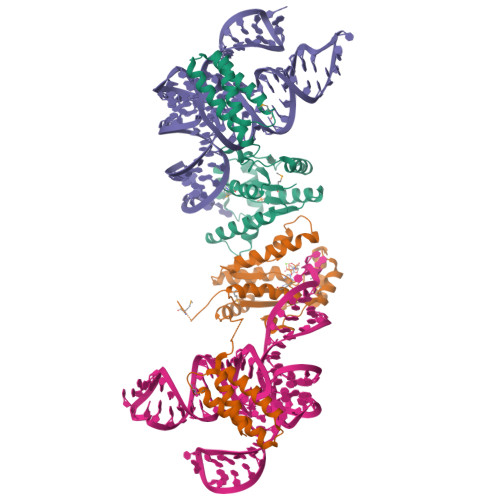
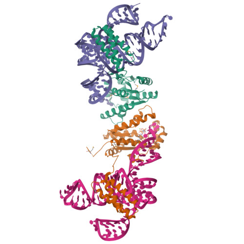
Crystal structure of O-phosphoseryl-tRNA kinase complexed with selenocysteine tRNA and AMPPNP (crystal type 1)


Experimental Data Snapshot

  • Method: X-RAY DIFFRACTION
  • Resolution: 2.80 Å
  • R-Value Free: 0.269 
  • R-Value Work: 0.228 
  • R-Value Observed: 0.230 

wwPDB Validation   3D Report Full Report


Ligand Structure Quality Assessment 


This is version 1.2 of the entry. See complete history


Literature

Structural Basis for the Major Role of O-Phosphoseryl-tRNA Kinase in the UGA-Specific Encoding of Selenocysteine

Chiba, S.Itoh, Y.Sekine, S.Yokoyama, S.

(2010) Mol Cell 39: 410-420

  • DOI: https://doi.org/10.1016/j.molcel.2010.07.018
  • Primary Citation of Related Structures:  
    3ADB, 3ADC, 3ADD

  • PubMed Abstract: 

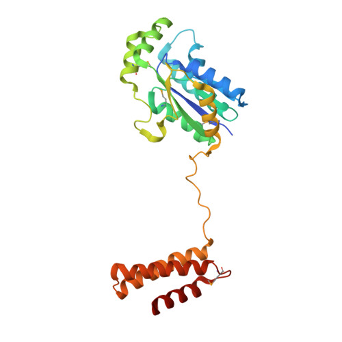
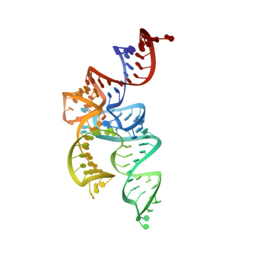
    The 21(st) amino acid, selenocysteine (Sec), is assigned to the codon UGA and is biosynthesized on the selenocysteine-specific tRNA (tRNA(Sec)) with the corresponding anticodon. In archaea/eukarya, tRNA(Sec) is ligated with serine by seryl-tRNA synthetase (SerRS), the seryl moiety is phosphorylated by O-phosphoseryl-tRNA kinase (PSTK), and the phosphate group is replaced with selenol by Sep-tRNA:Sec-tRNA synthase. PSTK selectively phosphorylates seryl-tRNA(Sec), while SerRS serylates both tRNA(Ser) and tRNA(Sec). In this study, we determined the crystal structures of the archaeal tRNA(Sec).PSTK complex. PSTK consists of two independent linker-connected domains, the N-terminal catalytic domain (NTD) and the C-terminal domain (CTD). The D-arm.CTD binding occurs independently of and much more strongly than the acceptor-arm.NTD binding. PSTK thereby distinguishes the characteristic D arm with the maximal stem and the minimal loop of tRNA(Sec) from the canonical D arm of tRNA(Ser), without interacting with the anticodon. This mechanism is essential for the UGA-specific encoding of selenocysteine.


  • Organizational Affiliation

    Department of Biophysics and Biochemistry, Graduate School of Science, The University of Tokyo, Tokyo, Japan.


Macromolecules

Find similar proteins by:  (by identity cutoff)  |  3D Structure
Entity ID: 1
MoleculeChains Sequence LengthOrganismDetailsImage
L-seryl-tRNA(Sec) kinase
A, B
259Methanocaldococcus jannaschii DSM 2661Mutation(s): 0 
Gene Names: MJ1538
EC: 2.7.1 (PDB Primary Data), 2.7.1.164 (UniProt)
UniProt
Find proteins for Q58933 (Methanocaldococcus jannaschii (strain ATCC 43067 / DSM 2661 / JAL-1 / JCM 10045 / NBRC 100440))
Explore Q58933 
Go to UniProtKB:  Q58933
Entity Groups  
Sequence Clusters30% Identity50% Identity70% Identity90% Identity95% Identity100% Identity
UniProt GroupQ58933
Sequence Annotations
Expand
  • Reference Sequence
Find similar nucleic acids by:  (by identity cutoff)  |  3D Structure
Entity ID: 2
MoleculeChains LengthOrganismImage
selenocysteine tRNA
C, D
92Methanopyrus kandleri
Sequence Annotations
Expand
  • Reference Sequence
Small Molecules
Ligands 3 Unique
IDChains Name / Formula / InChI Key2D Diagram3D Interactions
ANP
Query on ANP

Download Ideal Coordinates CCD File 
F [auth B]PHOSPHOAMINOPHOSPHONIC ACID-ADENYLATE ESTER
C10 H17 N6 O12 P3
PVKSNHVPLWYQGJ-KQYNXXCUSA-N
PO4
Query on PO4

Download Ideal Coordinates CCD File 
E [auth A]PHOSPHATE ION
O4 P
NBIIXXVUZAFLBC-UHFFFAOYSA-K
MG
Query on MG

Download Ideal Coordinates CCD File 
G [auth B]MAGNESIUM ION
Mg
JLVVSXFLKOJNIY-UHFFFAOYSA-N
Modified Residues  1 Unique
IDChains TypeFormula2D DiagramParent
MSE
Query on MSE
A, B
L-PEPTIDE LINKINGC5 H11 N O2 SeMET
Experimental Data & Validation

Experimental Data

  • Method: X-RAY DIFFRACTION
  • Resolution: 2.80 Å
  • R-Value Free: 0.269 
  • R-Value Work: 0.228 
  • R-Value Observed: 0.230 
  • Space Group: P 21 21 2
Unit Cell:
Length ( Å )Angle ( ˚ )
a = 92.196α = 90
b = 260.923β = 90
c = 45.558γ = 90
Software Package:
Software NamePurpose
HKL-2000data collection
autoSHARPphasing
CNSrefinement
HKL-2000data reduction
HKL-2000data scaling

Structure Validation

View Full Validation Report


Ligand Structure Quality Assessment 


Entry History 

Deposition Data

Revision History  (Full details and data files)

  • Version 1.0: 2010-07-28
    Type: Initial release
  • Version 1.1: 2011-07-13
    Changes: Version format compliance
  • Version 1.2: 2024-10-09
    Changes: Data collection, Database references, Derived calculations, Structure summary