3ZYM | pdb_00003zym

Structure of CALM (PICALM) in complex with VAMP8


Experimental Data Snapshot

  • Method: X-RAY DIFFRACTION
  • Resolution: 2.03 Å
  • R-Value Free: 
    0.198 (Depositor), 0.194 (DCC) 
  • R-Value Work: 
    0.181 (Depositor), 0.175 (DCC) 
  • R-Value Observed: 
    0.181 (Depositor) 

Starting Model: experimental
View more details

wwPDB Validation 3D Report Full Report

Validation slider image for 3ZYM

This is version 1.3 of the entry. See complete history

Literature

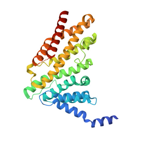
The molecular basis for the endocytosis of small R-SNAREs by the clathrin adaptor CALM.

Miller, S.E.Sahlender, D.A.Graham, S.C.Honing, S.Robinson, M.S.Peden, A.A.Owen, D.J.

(2011) Cell 147: 1118-1131

  • DOI: https://doi.org/10.1016/j.cell.2011.10.038
  • Primary Citation Related Structures: 
    3ZYK, 3ZYL, 3ZYM

  • PubMed Abstract: 

    SNAREs provide a large part of the specificity and energy needed for membrane fusion and, to do so, must be localized to their correct membranes. Here, we show that the R-SNAREs VAMP8, VAMP3, and VAMP2, which cycle between the plasma membrane and endosomes, bind directly to the ubiquitously expressed, PtdIns4,5P(2)-binding, endocytic clathrin adaptor CALM/PICALM. X-ray crystallography shows that the N-terminal halves of their SNARE motifs bind the CALM(ANTH) domain as helices in a manner that mimics SNARE complex formation. Mutation of residues in the CALM:SNARE interface inhibits binding in vitro and prevents R-SNARE endocytosis in vivo. Thus, CALM:R-SNARE interactions ensure that R-SNAREs, required for the fusion of endocytic clathrin-coated vesicles with endosomes and also for subsequent postendosomal trafficking, are sorted into endocytic vesicles. CALM's role in directing the endocytosis of small R-SNAREs may provide insight into the association of CALM/PICALM mutations with growth retardation, cognitive defects, and Alzheimer's disease.


  • Organizational Affiliation
    • Cambridge Institute for Medical Research and Department of Clinical Biochemistry, University of Cambridge, Addenbrooke's Hospital, Hills Road, Cambridge CB2 0XY, UK.

Macromolecule Content 

  • Total Structure Weight: 106.59 kDa 
  • Atom Count: 7,357 
  • Modeled Residue Count: 836 
  • Deposited Residue Count: 930 
  • Unique protein chains: 1

Macromolecules

Find similar proteins by:|  3D Structure
Entity ID: 1
MoleculeChains  Sequence LengthOrganismDetailsImage
PHOSPHATIDYLINOSITOL-BINDING CLATHRIN ASSEMBLY PROTEIN, VESICLE-ASSOCIATED MEMBRANE PROTEIN 8
A, B, C
310Rattus norvegicusMus musculus
This entity is chimeric
Mutation(s): 0 
UniProt
Find proteins for O55012 (Rattus norvegicus)
Explore O55012 
Go to UniProtKB:  O55012
Find proteins for O70404 (Mus musculus)
Explore O70404 
Go to UniProtKB:  O70404
Entity Groups
Sequence Clusters30% Identity50% Identity70% Identity90% Identity95% Identity100% Identity
UniProt GroupsO55012O70404
Sequence Annotations
Expand
Reference Sequence

Small Molecules

Ligands 2 Unique
IDChains Name / Formula / InChI Key2D Diagram3D Interactions
PO4

Query on PO4


Download:Ideal Coordinates CCD File
F [auth A]
G [auth A]
H [auth A]
K [auth B]
L [auth B]
F [auth A],
G [auth A],
H [auth A],
K [auth B],
L [auth B],
O [auth C],
P [auth C]
PHOSPHATE ION
O4 P
NBIIXXVUZAFLBC-UHFFFAOYSA-K
GOL

Query on GOL


Download:Ideal Coordinates CCD File
D [auth A]
E [auth A]
I [auth B]
J [auth B]
M [auth C]
D [auth A],
E [auth A],
I [auth B],
J [auth B],
M [auth C],
N [auth C]
GLYCEROL
C3 H8 O3
PEDCQBHIVMGVHV-UHFFFAOYSA-N

Experimental Data & Validation

Experimental Data

  • Method: X-RAY DIFFRACTION
  • Resolution: 2.03 Å
  • R-Value Free:  0.198 (Depositor), 0.194 (DCC) 
  • R-Value Work:  0.181 (Depositor), 0.175 (DCC) 
  • R-Value Observed: 0.181 (Depositor) 
Space Group: C 1 2 1
Unit Cell:
Length ( Å )Angle ( ˚ )
a = 161.051α = 90
b = 100.26β = 118.88
c = 104.021γ = 90
Software Package:
Software NamePurpose
PHENIXrefinement
MOSFLMdata reduction
SCALAdata scaling
PHASERphasing

Structure Validation

View Full Validation Report


Entry History 

Deposition Data

Revision History  (Full details and data files)

  • Version 1.0: 2011-12-07
    Type: Initial release
  • Version 1.1: 2017-03-15
    Changes: Source and taxonomy
  • Version 1.2: 2019-03-13
    Changes: Data collection, Database references, Experimental preparation
  • Version 1.3: 2023-12-20
    Changes: Data collection, Database references, Derived calculations, Other, Refinement description