3ZXX | pdb_00003zxx

Structure of self-cleaved protease domain of PatA


Experimental Data Snapshot

  • Method: X-RAY DIFFRACTION
  • Resolution: 1.95 Å
  • R-Value Free: 
    0.174 (Depositor), 0.182 (DCC) 
  • R-Value Work: 
    0.137 (Depositor), 0.147 (DCC) 
  • R-Value Observed: 
    0.139 (Depositor) 

Starting Model: experimental
View more details

wwPDB Validation 3D Report Full Report

Validation slider image for 3ZXX

This is version 1.5 of the entry. See complete history

Literature

The Discovery of New Cyanobactins from Cyanothece Pcc 7425 Defines a New Signature for Processing of Patellamides.

Houssen, W.E.Koehnke, J.Zollman, D.Vendome, J.Raab, A.Smith, M.C.Naismith, J.H.Jaspars, M.

(2012) Chembiochem 13: 2683

  • DOI: https://doi.org/10.1002/cbic.201200661
  • Primary Citation Related Structures: 
    3ZXX, 3ZXY

  • PubMed Abstract: 

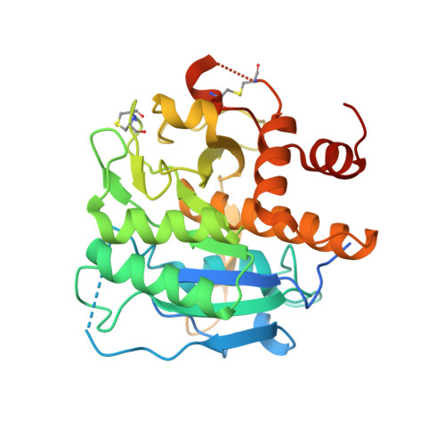
    Cyanobactins, including patellamides, are a group of cyanobacterial post-translationally modified ribosomal cyclic peptides. The final product should in theory be predictable from the sequence of the precursor peptide and the associated tailoring enzymes. Understanding the mechanism and recognition requirements of these enzymes will allow their rational engineering. We have identified three new cyanobactins from a Cyanothece PCC 7425 culture subjected to a heat shock. One of these compounds revealed a novel signature signal for ThcA, the subtilisin-like serine protease that is homologous to the patellamide protease PatA. The crystal structure of the latter and modelling studies allow rationalisation of the recognition determinants for both enzymes, consistent with the ribosomal biosynthetic origin of the new compounds.


  • Organizational Affiliation
    • Marine Biodiscovery Centre, Department of Chemistry, University of Aberdeen, UK.

Macromolecule Content 

  • Total Structure Weight: 32.37 kDa 
  • Atom Count: 2,341 
  • Modeled Residue Count: 265 
  • Deposited Residue Count: 307 
  • Unique protein chains: 1

Macromolecules

Find similar proteins by:|  3D Structure
Entity ID: 1
MoleculeChains  Sequence LengthOrganismDetailsImage
SUBTILISIN-LIKE PROTEIN307Prochloron didemniMutation(s): 0 
UniProt
Find proteins for Q52QI9 (Prochloron didemni)
Explore Q52QI9 
Go to UniProtKB:  Q52QI9
Entity Groups
Sequence Clusters30% Identity50% Identity70% Identity90% Identity95% Identity100% Identity
UniProt GroupQ52QI9
Sequence Annotations
Expand
Reference Sequence

Experimental Data & Validation

Experimental Data

  • Method: X-RAY DIFFRACTION
  • Resolution: 1.95 Å
  • R-Value Free:  0.174 (Depositor), 0.182 (DCC) 
  • R-Value Work:  0.137 (Depositor), 0.147 (DCC) 
  • R-Value Observed: 0.139 (Depositor) 
Space Group: P 41
Unit Cell:
Length ( Å )Angle ( ˚ )
a = 84.21α = 90
b = 84.21β = 90
c = 41.84γ = 90
Software Package:
Software NamePurpose
REFMACrefinement
PHASERphasing

Structure Validation

View Full Validation Report


Entry History 

Deposition Data

Revision History  (Full details and data files)

  • Version 1.0: 2012-08-29
    Type: Initial release
  • Version 1.1: 2012-12-05
    Changes: Database references
  • Version 1.2: 2013-01-16
    Changes: Database references
  • Version 1.3: 2013-11-06
    Changes: Structure summary
  • Version 1.4: 2023-12-20
    Changes: Data collection, Database references, Other, Refinement description
  • Version 1.5: 2024-11-13
    Changes: Structure summary