3ZWS | pdb_00003zws

Structure of Human Dihydroorotate Dehydrogenase with a Bound Inhibitor


Experimental Data Snapshot

  • Method: X-RAY DIFFRACTION
  • Resolution: 1.60 Å
  • R-Value Free: 
    0.176 (Depositor), 0.185 (DCC) 
  • R-Value Work: 
    0.161 (Depositor) 
  • R-Value Observed: 
    0.162 (Depositor) 

wwPDB Validation 3D Report Full Report

Validation slider image for 3ZWS

Ligand Structure Quality Assessment 


This is version 1.2 of the entry. See complete history

Literature

Factors Influencing the Specificity of Inhibitor Binding to the Human and Malaria Parasite Dihydroorotate Dehydrogenases.

Bedingfield, P.T.P.Cowen, D.Acklam, P.A.Cunningham, F.Parsons, M.R.Mcconkey, G.A.Fishwick, C.W.G.Johnson, A.P.

(2012) J Med Chem 55: 5841

  • DOI: https://doi.org/10.1021/jm300157n
  • Primary Citation Related Structures: 
    3ZWS, 3ZWT

  • PubMed Abstract: 

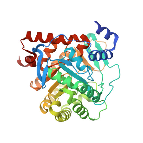
    The de novo pyrimidine biosynthesis enzyme dihydroorotate dehydrogenase is an emerging drug target for the treatment of malaria. In this context a key property of Plasmodium falciparum DHODH (PfDHODH) is that it can be selectively inhibited over its human homologue (HsDHODH). However, HsDHODH is also a validated drug target for autoimmune diseases such as arthritis. Here a series of novel inhibitors is described that includes compounds that switch specificity between the two enzymes as a result of small alterations in chemical structure. Structure-activity relationship (SAR), crystallography, docking, and mutagenesis studies are used to examine the binding modes of the compounds within the two enzymes and to reveal structural changes induced by inhibitor binding. Within this series, compounds with therapeutically relevant HsDHODH activity are described and their binding modes characterized using X-ray crystallography, which reveals a novel conformational shift within the inhibitor binding site.


  • Organizational Affiliation
    • Faculty of Biological Sciences, University of Leeds, Leeds, LS2 9JT, U.K.

Macromolecule Content 

  • Total Structure Weight: 41.16 kDa 
  • Atom Count: 3,097 
  • Modeled Residue Count: 354 
  • Deposited Residue Count: 367 
  • Unique protein chains: 1

Macromolecules

Find similar proteins by:|  3D Structure
Entity ID: 1
MoleculeChains  Sequence LengthOrganismDetailsImage
DIHYDROOROTATE DEHYDROGENASE (QUINONE), MITOCHONDRIAL367Homo sapiensMutation(s): 0 
EC: 1.3.5.2 (PDB Primary Data), 1.3.3.1 (PDB Primary Data)
UniProt & NIH Common Fund Data Resources
Find proteins for Q02127 (Homo sapiens)
Explore Q02127 
Go to UniProtKB:  Q02127
PHAROS:  Q02127
GTEx:  ENSG00000102967 
Entity Groups
Sequence Clusters30% Identity50% Identity70% Identity90% Identity95% Identity100% Identity
UniProt GroupQ02127
Sequence Annotations
Expand
Reference Sequence

Small Molecules

Ligands 6 Unique
IDChains Name / Formula / InChI Key2D Diagram3D Interactions
FMN

Query on FMN


Download:Ideal Coordinates CCD File
B [auth A]FLAVIN MONONUCLEOTIDE
C17 H21 N4 O9 P
FVTCRASFADXXNN-SCRDCRAPSA-N
AVQ

Query on AVQ


Download:Ideal Coordinates CCD File
G [auth A]2-[(2,5-DICHLOROBENZYL)SULFANYL]-5-METHYL[1,2,4]TRIAZOLO[1,5-A]PYRIMIDIN-7-OL
C13 H10 Cl2 N4 O S
VQASKKWJQGYGRL-UHFFFAOYSA-N
ORO

Query on ORO


Download:Ideal Coordinates CCD File
C [auth A]OROTIC ACID
C5 H4 N2 O4
PXQPEWDEAKTCGB-UHFFFAOYSA-N
SO4

Query on SO4


Download:Ideal Coordinates CCD File
D [auth A],
E [auth A]
SULFATE ION
O4 S
QAOWNCQODCNURD-UHFFFAOYSA-L
ACT

Query on ACT


Download:Ideal Coordinates CCD File
H [auth A],
I [auth A]
ACETATE ION
C2 H3 O2
QTBSBXVTEAMEQO-UHFFFAOYSA-M
CL

Query on CL


Download:Ideal Coordinates CCD File
F [auth A]CHLORIDE ION
Cl
VEXZGXHMUGYJMC-UHFFFAOYSA-M

Experimental Data & Validation

Experimental Data

  • Method: X-RAY DIFFRACTION
  • Resolution: 1.60 Å
  • R-Value Free:  0.176 (Depositor), 0.185 (DCC) 
  • R-Value Work:  0.161 (Depositor) 
  • R-Value Observed: 0.162 (Depositor) 
Space Group: P 32 2 1
Unit Cell:
Length ( Å )Angle ( ˚ )
a = 90.72α = 90
b = 90.72β = 90
c = 123.072γ = 120
Software Package:
Software NamePurpose
REFMACrefinement
MOSFLMdata reduction
SCALAdata scaling

Structure Validation

View Full Validation Report


Ligand Structure Quality Assessment 


Entry History 

Deposition Data

Revision History  (Full details and data files)

  • Version 1.0: 2012-06-27
    Type: Initial release
  • Version 1.1: 2012-07-11
    Changes: Other
  • Version 1.2: 2024-05-08
    Changes: Data collection, Database references, Derived calculations, Other