3ZU5 | pdb_00003zu5

Structure of the enoyl-ACP reductase FabV from Yersinia pestis with the cofactor NADH and the 2-pyridone inhibitor PT173


Experimental Data Snapshot

  • Method: X-RAY DIFFRACTION
  • Resolution: 2.00 Å
  • R-Value Free: 
    0.228 (Depositor), 0.241 (DCC) 
  • R-Value Work: 
    0.187 (Depositor), 0.199 (DCC) 
  • R-Value Observed: 
    0.189 (Depositor) 

wwPDB Validation 3D Report Full Report

Validation slider image for 3ZU5

Ligand Structure Quality Assessment 


This is version 1.3 of the entry. See complete history

Literature

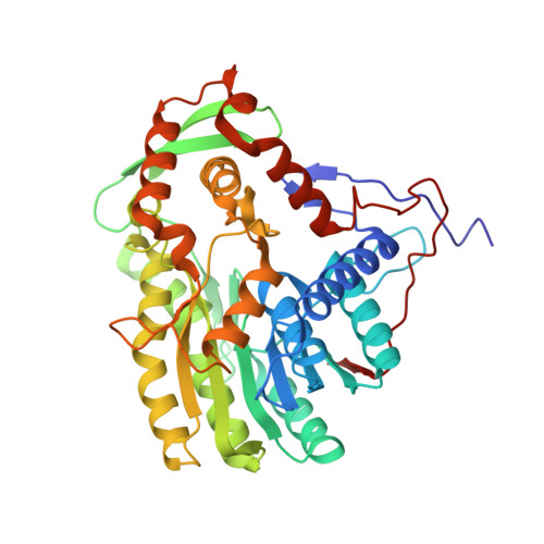
Structure of the Yersinia Pestis Fabv Enoyl-Acp Reductase and its Interaction with Two 2-Pyridone Inhibitors

Hirschbeck, M.W.Kuper, J.Lu, H.Liu, N.Neckles, C.Shah, S.Wagner, S.Sotriffer, C.A.Tonge, P.J.Kisker, C.

(2012) Structure 20: 89

  • DOI: https://doi.org/10.1016/j.str.2011.07.019
  • Primary Citation Related Structures: 
    3ZU2, 3ZU3, 3ZU4, 3ZU5

  • PubMed Abstract: 

    The recently discovered FabV enoyl-ACP reductase, which catalyzes the last step of the bacterial fatty acid biosynthesis (FAS-II) pathway, is a promising but unexploited drug target against the reemerging pathogen Yersinia pestis. The structure of Y. pestis FabV in complex with its cofactor reveals that the enzyme features the common architecture of the short-chain dehydrogenase reductase superfamily, but contains additional structural elements that are mostly folded around the usually flexible substrate-binding loop, thereby stabilizing it in a very tight conformation that seals the active site. The structures of FabV in complex with NADH and two newly developed 2-pyridone inhibitors provide insights for the development of new lead compounds, and suggest a mechanism by which the substrate-binding loop opens to admit the inhibitor, a motion that could also be coupled to the interaction of FabV with the acyl-carrier protein substrate.


  • Organizational Affiliation
    • Rudolf Virchow Center for Experimental Biomedicine, Institute for Structural Biology, Josef-Schneider-Strasse 2, University of Würzburg, D-97080 Würzburg, Germany.

Macromolecule Content 

  • Total Structure Weight: 45.02 kDa 
  • Atom Count: 3,569 
  • Modeled Residue Count: 404 
  • Deposited Residue Count: 405 
  • Unique protein chains: 1

Macromolecules

Find similar proteins by:|  3D Structure
Entity ID: 1
MoleculeChains  Sequence LengthOrganismDetailsImage
PUTATIVE REDUCTASE YPO4104/Y4119/YP_4011405Yersinia pestis CO92Mutation(s): 1 
EC: 1.3.1 (PDB Primary Data), 1.3.1.9 (UniProt)
UniProt
Find proteins for Q8Z9U1 (Yersinia pestis)
Explore Q8Z9U1 
Go to UniProtKB:  Q8Z9U1
Entity Groups
Sequence Clusters30% Identity50% Identity70% Identity90% Identity95% Identity100% Identity
UniProt GroupQ8Z9U1
Sequence Annotations
Expand
Reference Sequence

Small Molecules

Binding Affinity Annotations 
IDSourceBinding Affinity
AEW BindingDB:  3ZU5 IC50: min: 1000, max: 1.00e+5 (nM) from 2 assay(s)

Experimental Data & Validation

Experimental Data

  • Method: X-RAY DIFFRACTION
  • Resolution: 2.00 Å
  • R-Value Free:  0.228 (Depositor), 0.241 (DCC) 
  • R-Value Work:  0.187 (Depositor), 0.199 (DCC) 
  • R-Value Observed: 0.189 (Depositor) 
Space Group: P 31 2 1
Unit Cell:
Length ( Å )Angle ( ˚ )
a = 101.77α = 90
b = 101.77β = 90
c = 84.55γ = 120
Software Package:
Software NamePurpose
REFMACrefinement
iMOSFLMdata reduction
SCALAdata scaling
PHASERphasing

Structure Validation

View Full Validation Report


Ligand Structure Quality Assessment 


Entry History 

Deposition Data

Revision History  (Full details and data files)

  • Version 1.0: 2012-01-18
    Type: Initial release
  • Version 1.1: 2012-03-28
    Changes: Other
  • Version 1.2: 2017-07-05
    Changes: Data collection
  • Version 1.3: 2024-05-08
    Changes: Data collection, Database references, Derived calculations, Other