3ZT4 | pdb_00003zt4

Small molecule inhibitors of the LEDGF site of HIV type 1 integrase identified by fragment screening and structure based drug design


Experimental Data Snapshot

  • Method: X-RAY DIFFRACTION
  • Resolution: 2.20 Å
  • R-Value Free: 
    0.229 (Depositor), 0.208 (DCC) 
  • R-Value Work: 
    0.170 (Depositor) 
  • R-Value Observed: 
    0.173 (Depositor) 

Starting Model: experimental
View more details

wwPDB Validation 3D Report Full Report

Validation slider image for 3ZT4

Ligand Structure Quality Assessment 


This is version 1.2 of the entry. See complete history

Literature

Small Molecule Inhibitors of the Ledgf Site of Human Immunodeficiency Virus Integrase Identified by Fragment Screening and Structure Based Design

Peat, T.S.Rhodes, D.I.Vandergraaff, N.Le, G.Smith, J.A.Clark, L.J.Jones, E.D.Coates, J.A.V.Thienthong, N.Newman, J.Dolezal, O.Mulder, R.Ryan, J.H.Savage, G.P.Francis, C.L.Deadman, J.J.

(2012) PLoS One 7: 40147

  • DOI: https://doi.org/10.1371/journal.pone.0040147
  • Primary Citation Related Structures: 
    3ZCM, 3ZSO, 3ZSQ, 3ZSR, 3ZSV, 3ZSW, 3ZSX, 3ZSY, 3ZSZ, 3ZT0, 3ZT1, 3ZT2, 3ZT3, 3ZT4

  • PubMed Abstract: 

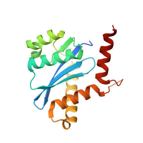
    A fragment-based screen against human immunodeficiency virus type 1 (HIV) integrase led to a number of compounds that bound to the lens epithelium derived growth factor (LEDGF) binding site of the integrase catalytic core domain. We determined the crystallographic structures of complexes of the HIV integrase catalytic core domain for 10 of these compounds and quantitated the binding by surface plasmon resonance. We demonstrate that the compounds inhibit the interaction of LEDGF with HIV integrase in a proximity AlphaScreen assay, an assay for the LEDGF enhancement of HIV integrase strand transfer and in a cell based assay. The compounds identified represent a potential framework for the development of a new series of HIV integrase inhibitors that do not bind to the catalytic site of the enzyme.


  • Organizational Affiliation
    • CSIRO Materials, Science and Engineering, Parkville, Victoria, Australia. tom.peat@csiro.au

Macromolecule Content 

  • Total Structure Weight: 38.78 kDa 
  • Atom Count: 2,670 
  • Modeled Residue Count: 301 
  • Deposited Residue Count: 334 
  • Unique protein chains: 1

Macromolecules

Find similar proteins by:|  3D Structure
Entity ID: 1
MoleculeChains  Sequence LengthOrganismDetailsImage
INTEGRASE
A, B
167Human immunodeficiency virusMutation(s): 3 
EC: 2.7.7
UniProt
Find proteins for P12497 (Human immunodeficiency virus type 1 group M subtype B (isolate NY5))
Explore P12497 
Go to UniProtKB:  P12497
Entity Groups
Sequence Clusters30% Identity50% Identity70% Identity90% Identity95% Identity100% Identity
UniProt GroupP12497
Sequence Annotations
Expand
Reference Sequence

Small Molecules

Ligands 4 Unique
IDChains Name / Formula / InChI Key2D Diagram3D Interactions
ZT2

Query on ZT2


Download:Ideal Coordinates CCD File
H [auth A],
I [auth A],
N [auth B],
O [auth B]
5-[(E)-(2-OXO-2,3-DIHYDRO-1H-INDEN-1-YLIDENE)METHYL]-1,3-BENZODIOXOLE-4-CARBOXYLIC ACID
C18 H12 O5
WRNAEMYUELGDPO-NTUHNPAUSA-N
SO4

Query on SO4


Download:Ideal Coordinates CCD File
C [auth A]
D [auth A]
E [auth A]
J [auth B]
K [auth B]
C [auth A],
D [auth A],
E [auth A],
J [auth B],
K [auth B],
L [auth B]
SULFATE ION
O4 S
QAOWNCQODCNURD-UHFFFAOYSA-L
EDO

Query on EDO


Download:Ideal Coordinates CCD File
G [auth A]1,2-ETHANEDIOL
C2 H6 O2
LYCAIKOWRPUZTN-UHFFFAOYSA-N
ACY

Query on ACY


Download:Ideal Coordinates CCD File
F [auth A],
M [auth B]
ACETIC ACID
C2 H4 O2
QTBSBXVTEAMEQO-UHFFFAOYSA-N

Experimental Data & Validation

Experimental Data

  • Method: X-RAY DIFFRACTION
  • Resolution: 2.20 Å
  • R-Value Free:  0.229 (Depositor), 0.208 (DCC) 
  • R-Value Work:  0.170 (Depositor) 
  • R-Value Observed: 0.173 (Depositor) 
Space Group: P 31
Unit Cell:
Length ( Å )Angle ( ˚ )
a = 71.058α = 90
b = 71.058β = 90
c = 66.559γ = 120
Software Package:
Software NamePurpose
REFMACrefinement
MOSFLMdata reduction
SCALAdata scaling
PHASERphasing

Structure Validation

View Full Validation Report


Ligand Structure Quality Assessment 


Entry History 

Revision History  (Full details and data files)

  • Version 1.0: 2012-07-18
    Type: Initial release
  • Version 1.1: 2012-08-01
    Changes: Database references
  • Version 1.2: 2023-12-20
    Changes: Data collection, Database references, Derived calculations, Other, Refinement description