3ZRX

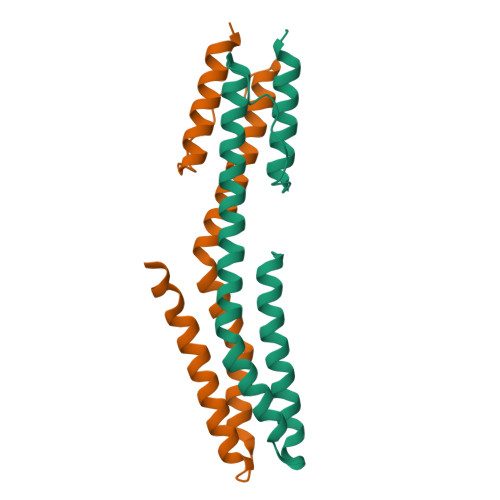
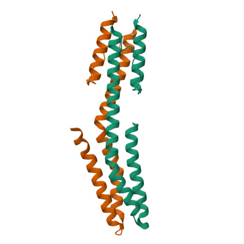
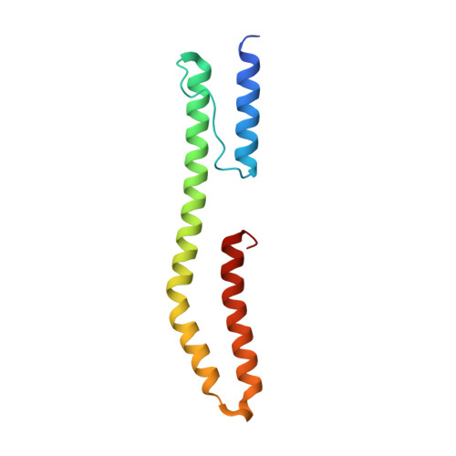
The high resolution structure of a dimeric Hamp-Dhp fusion displays strong asymmetry


Experimental Data Snapshot

  • Method: X-RAY DIFFRACTION
  • Resolution: 1.25 Å
  • R-Value Free: 0.200 
  • R-Value Work: 0.151 
  • R-Value Observed: 0.153 

Starting Model: experimental
View more details

wwPDB Validation   3D Report Full Report


This is version 1.3 of the entry. See complete history


Literature

Mechanism of Regulation of Receptor Histidine Kinases.

Ferris, H.U.Dunin-Horkawicz, S.Hornig, N.Hulko, M.Martin, J.Schultz, J.E.Zeth, K.Lupas, A.N.Coles, M.

(2012) Structure 20: 56

  • DOI: https://doi.org/10.1016/j.str.2011.11.014
  • Primary Citation of Related Structures:  
    2LFR, 2LFS, 3ZRV, 3ZRW, 3ZRX

  • PubMed Abstract: 

    Bacterial transmembrane receptors regulate an intracellular catalytic output in response to extracellular sensory input. To investigate the conformational changes that relay the regulatory signal, we have studied the HAMP domain, a ubiquitous intracellular module connecting input to output domains. HAMP forms a parallel, dimeric, four-helical coiled coil, and rational substitutions in our model domain (Af1503 HAMP) induce a transition in its interhelical packing, characterized by axial rotation of all four helices (the gearbox signaling model). We now illustrate how these conformational changes are propagated to a downstream domain by fusing Af1503 HAMP variants to the DHp domain of EnvZ, a bacterial histidine kinase. Structures of wild-type and mutant constructs are correlated with ligand response in vivo, clearly associating them with distinct signaling states. We propose that altered recognition of the catalytic domain by DHp, rather than a shift in position of the phospho-accepting histidine, forms the basis for regulation of kinase activity.


  • Organizational Affiliation

    Department of Protein Evolution, Max-Planck-Institute for Developmental Biology, 72076 Tübingen, Germany.


Macromolecules
Find similar proteins by:  (by identity cutoff)  |  3D Structure
Entity ID: 1
MoleculeChains Sequence LengthOrganismDetailsImage
AF1503 PROTEIN, OSMOLARITY SENSOR PROTEIN ENVZ
A, B
115Archaeoglobus fulgidus DSM 4304Escherichia coli K-12
This entity is chimeric
Mutation(s): 0 
EC: 2.7.13.3
UniProt
Find proteins for P0AEJ4 (Escherichia coli (strain K12))
Explore P0AEJ4 
Go to UniProtKB:  P0AEJ4
Find proteins for O28769 (Archaeoglobus fulgidus (strain ATCC 49558 / DSM 4304 / JCM 9628 / NBRC 100126 / VC-16))
Explore O28769 
Go to UniProtKB:  O28769
Entity Groups  
Sequence Clusters30% Identity50% Identity70% Identity90% Identity95% Identity100% Identity
UniProt GroupsP0AEJ4O28769
Sequence Annotations
Expand
  • Reference Sequence
Experimental Data & Validation

Experimental Data

  • Method: X-RAY DIFFRACTION
  • Resolution: 1.25 Å
  • R-Value Free: 0.200 
  • R-Value Work: 0.151 
  • R-Value Observed: 0.153 
  • Space Group: P 1 21 1
Unit Cell:
Length ( Å )Angle ( ˚ )
a = 36.11α = 90
b = 57.4β = 107.32
c = 51.62γ = 90
Software Package:
Software NamePurpose
REFMACrefinement
XDSdata reduction
XSCALEdata scaling
SHARPphasing
DMphasing
BUCCANEERmodel building

Structure Validation

View Full Validation Report


Entry History 

Deposition Data

Revision History  (Full details and data files)

  • Version 1.0: 2011-06-29
    Type: Initial release
  • Version 1.1: 2012-04-25
    Changes: Database references, Version format compliance
  • Version 1.2: 2017-03-15
    Changes: Source and taxonomy
  • Version 1.3: 2023-12-20
    Changes: Data collection, Database references, Other, Refinement description