3ZPZ | pdb_00003zpz

Visualizing GroEL-ES in the Act of Encapsulating a Non-Native Substrate Protein


Experimental Data Snapshot

  • Method: ELECTRON MICROSCOPY
  • Resolution: 8.90 Å
  • Aggregation State: PARTICLE 
  • Reconstruction Method: SINGLE PARTICLE 

Starting Model: experimental
View more details

wwPDB Validation 3D Report Full Report

Validation slider image for 3ZPZ

This is version 1.2 of the entry. See complete history

Literature

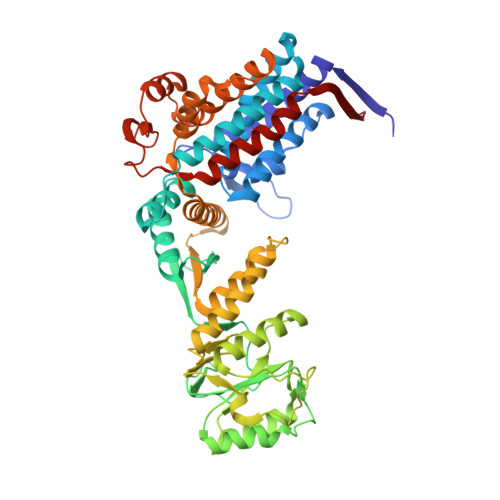
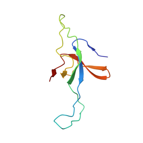
Visualizing Groel/Es in the Act of Encapsulating a Folding Protein

Chen, D.-H.Madan, D.Weaver, J.Lin, Z.Schroder, G.F.Chiu, W.Rye, H.S.

(2013) Cell 153: 1354

  • DOI: https://doi.org/10.1016/j.cell.2013.04.052
  • Primary Citation Related Structures: 
    3ZPZ, 3ZQ0, 3ZQ1

  • PubMed Abstract: 

    The GroEL/ES chaperonin system is required for the assisted folding of many proteins. How these substrate proteins are encapsulated within the GroEL-GroES cavity is poorly understood. Using symmetry-free, single-particle cryo-electron microscopy, we have characterized a chemically modified mutant of GroEL (EL43Py) that is trapped at a normally transient stage of substrate protein encapsulation. We show that the symmetric pattern of the GroEL subunits is broken as the GroEL cis-ring apical domains reorient to accommodate the simultaneous binding of GroES and an incompletely folded substrate protein (RuBisCO). The collapsed RuBisCO folding intermediate binds to the lower segment of two apical domains, as well as to the normally unstructured GroEL C-terminal tails. A comparative structural analysis suggests that the allosteric transitions leading to substrate protein release and folding involve concerted shifts of GroES and the GroEL apical domains and C-terminal tails.


  • Organizational Affiliation
    • Verna and Marrs McLean Department of Biochemistry and Molecular Biology, National Center for Macromolecular Imaging, Baylor College of Medicine, Houston, TX 77030, USA.

Macromolecule Content 

  • Total Structure Weight: 852.45 kDa 
  • Atom Count: 59,276 
  • Modeled Residue Count: 8,029 
  • Deposited Residue Count: 8,043 
  • Unique protein chains: 2

Macromolecules

Find similar proteins by:|  3D Structure
Entity ID: 1
MoleculeChains  Sequence LengthOrganismDetailsImage
60 KDA CHAPERONIN
A, B, C, D, E
A, B, C, D, E, F, G, H, I, J, K, L, M, N
526Escherichia coli BL21Mutation(s): 0 
EC: 5.6.1.7
UniProt
Find proteins for P0A6F5 (Escherichia coli (strain K12))
Explore P0A6F5 
Go to UniProtKB:  P0A6F5
Entity Groups
Sequence Clusters30% Identity50% Identity70% Identity90% Identity95% Identity100% Identity
UniProt GroupP0A6F5
Sequence Annotations
Expand
Reference Sequence
Find similar proteins by:|  3D Structure
Entity ID: 2
MoleculeChains  Sequence LengthOrganismDetailsImage
10 KDA CHAPERONIN
O, P, Q, R, S
O, P, Q, R, S, T, U
97Escherichia coli K-12Mutation(s): 0 
UniProt
Find proteins for P0A6F9 (Escherichia coli (strain K12))
Explore P0A6F9 
Go to UniProtKB:  P0A6F9
Entity Groups
Sequence Clusters30% Identity50% Identity70% Identity90% Identity95% Identity100% Identity
UniProt GroupP0A6F9
Sequence Annotations
Expand
Reference Sequence

Small Molecules

Ligands 2 Unique
IDChains Name / Formula / InChI Key2D Diagram3D Interactions
ADP

Query on ADP


Download:Ideal Coordinates CCD File
AA [auth C]
CA [auth D]
EA [auth E]
GA [auth F]
IA [auth G]
AA [auth C],
CA [auth D],
EA [auth E],
GA [auth F],
IA [auth G],
W [auth A],
Y [auth B]
ADENOSINE-5'-DIPHOSPHATE
C10 H15 N5 O10 P2
XTWYTFMLZFPYCI-KQYNXXCUSA-N
MG

Query on MG


Download:Ideal Coordinates CCD File
BA [auth D]
DA [auth E]
FA [auth F]
HA [auth G]
V [auth A]
BA [auth D],
DA [auth E],
FA [auth F],
HA [auth G],
V [auth A],
X [auth B],
Z [auth C]
MAGNESIUM ION
Mg
JLVVSXFLKOJNIY-UHFFFAOYSA-N

Experimental Data & Validation

Experimental Data

  • Method: ELECTRON MICROSCOPY
  • Resolution: 8.90 Å
  • Aggregation State: PARTICLE 
  • Reconstruction Method: SINGLE PARTICLE 
EM Software:
TaskSoftware PackageVersion
RECONSTRUCTIONEMAN1

Structure Validation

View Full Validation Report


Entry History 

Deposition Data

Revision History  (Full details and data files)

  • Version 1.0: 2013-06-19
    Type: Initial release
  • Version 1.1: 2017-08-23
    Changes: Data collection, Refinement description
  • Version 1.2: 2024-05-08
    Changes: Data collection, Database references, Derived calculations, Refinement description