3ZMN | pdb_00003zmn

VP17, a capsid protein of bacteriophage P23-77


Experimental Data Snapshot

  • Method: X-RAY DIFFRACTION
  • Resolution: 2.26 Å
  • R-Value Free: 
    0.213 (Depositor), 0.220 (DCC) 
  • R-Value Work: 
    0.185 (Depositor), 0.187 (DCC) 
  • R-Value Observed: 
    0.187 (Depositor) 

wwPDB Validation 3D Report Full Report

Validation slider image for 3ZMN

This is version 1.4 of the entry. See complete history

Literature

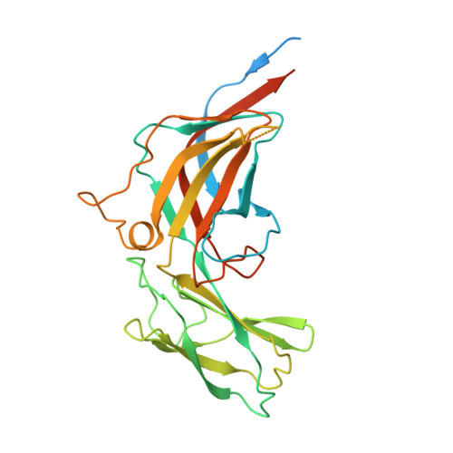
Bacteriophage P23-77 Capsid Protein Structures Reveal the Archetype of an Ancient Branch from a Major Virus Lineage.

Rissanen, I.Grimes, J.M.Pawlowski, A.Mantynen, S.Harlos, K.Bamford, J.K.H.Stuart, D.I.

(2013) Structure 21: 718

  • DOI: https://doi.org/10.1016/j.str.2013.02.026
  • Primary Citation Related Structures: 
    3ZMN, 3ZMO, 3ZN4, 3ZN5, 3ZN6

  • PubMed Abstract: 

    It has proved difficult to classify viruses unless they are closely related since their rapid evolution hinders detection of remote evolutionary relationships in their genetic sequences. However, structure varies more slowly than sequence, allowing deeper evolutionary relationships to be detected. Bacteriophage P23-77 is an example of a newly identified viral lineage, with members inhabiting extreme environments. We have solved multiple crystal structures of the major capsid proteins VP16 and VP17 of bacteriophage P23-77. They fit the 14 Å resolution cryo-electron microscopy reconstruction of the entire virus exquisitely well, allowing us to propose a model for both the capsid architecture and viral assembly, quite different from previously published models. The structures of the capsid proteins and their mode of association to form the viral capsid suggest that the P23-77-like and adeno-PRD1 lineages of viruses share an extremely ancient common ancestor.


  • Organizational Affiliation
    • Department of Biological and Environmental Science and Nanoscience Center, University of Jyväskylä, Jyväskylä 40014, Finland.

Macromolecule Content 

  • Total Structure Weight: 65.15 kDa 
  • Atom Count: 4,047 
  • Modeled Residue Count: 468 
  • Deposited Residue Count: 598 
  • Unique protein chains: 2

Macromolecules

Find similar proteins by:|  3D Structure
Entity ID: 1
MoleculeChains  Sequence LengthOrganismDetailsImage
VP17
A, B
291Thermus virus P23-77Mutation(s): 0 
UniProt
Find proteins for C8CHL5 (Thermus virus P23-77)
Explore C8CHL5 
Go to UniProtKB:  C8CHL5
Entity Groups
Sequence Clusters30% Identity50% Identity70% Identity90% Identity95% Identity100% Identity
UniProt GroupC8CHL5
Sequence Annotations
Expand
Reference Sequence
Find similar proteins by:  Sequence   |   3D Structure  
Entity ID: 2
MoleculeChains  Sequence LengthOrganismDetailsImage
VP17
C, D
8Thermus virus P23-77Mutation(s): 0 

Experimental Data & Validation

Experimental Data

  • Method: X-RAY DIFFRACTION
  • Resolution: 2.26 Å
  • R-Value Free:  0.213 (Depositor), 0.220 (DCC) 
  • R-Value Work:  0.185 (Depositor), 0.187 (DCC) 
  • R-Value Observed: 0.187 (Depositor) 
Space Group: P 61 2 2
Unit Cell:
Length ( Å )Angle ( ˚ )
a = 107.21α = 90
b = 107.21β = 90
c = 233.78γ = 120
Software Package:
Software NamePurpose
ARP/wARPmodel building
xia2data scaling
xia2phasing
SHELXDphasing
autoSHARPphasing
ARP/wARPphasing
BUSTERrefinement

Structure Validation

View Full Validation Report


Entry History 

Deposition Data

Revision History  (Full details and data files)

  • Version 1.0: 2013-05-15
    Type: Initial release
  • Version 1.1: 2013-05-22
    Changes: Atomic model, Database references, Non-polymer description, Other, Refinement description
  • Version 1.2: 2017-06-28
    Changes: Refinement description
  • Version 1.3: 2019-05-08
    Changes: Data collection, Experimental preparation
  • Version 1.4: 2024-05-08
    Changes: Data collection, Database references, Other