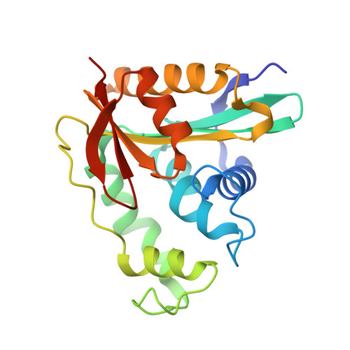
3ZJ0

The human O-GlcNAcase C-terminal domain is a pseudo histone acetyltransferase


Experimental Data Snapshot

  • Method: X-RAY DIFFRACTION
  • Resolution: 1.80 Å
  • R-Value Free: 0.234 
  • R-Value Work: 0.199 
  • R-Value Observed: 0.200 

wwPDB Validation   3D Report Full Report


Ligand Structure Quality Assessment 


This is version 1.1 of the entry. See complete history


Literature

Structure of a Bacterial Putative Acetyltransferase Defines the Fold of the Human O-Glcnacase C-Terminal Domain.

Rao, F.V.Schuttelkopf, A.W.Dorfmueller, H.C.Ferenbach, A.T.Navratilova, I.van Aalten, D.M.F.

(2013) Open Biol 3: 30021

  • DOI: https://doi.org/10.1098/rsob.130021
  • Primary Citation of Related Structures:  
    3ZJ0

  • PubMed Abstract: 

    The dynamic modification of proteins by O-linked N-acetylglucosamine (O-GlcNAc) is an essential posttranslational modification present in higher eukaryotes. Removal of O-GlcNAc is catalysed by O-GlcNAcase, a multi-domain enzyme that has been reported to be bifunctional, possessing both glycoside hydrolase and histone acetyltransferase (AT) activity. Insights into the mechanism, protein substrate recognition and inhibition of the hydrolase domain of human OGA (hOGA) have been obtained via the use of the structures of bacterial homologues. However, the molecular basis of AT activity of OGA, which has only been reported in vitro, is not presently understood. Here, we describe the crystal structure of a putative acetyltransferase (OgpAT) that we identified in the genome of the marine bacterium Oceanicola granulosus, showing homology to the hOGA C-terminal AT domain (hOGA-AT). The structure of OgpAT in complex with acetyl coenzyme A (AcCoA) reveals that, by homology modelling, hOGA-AT adopts a variant AT fold with a unique loop creating a deep tunnel. The structures, together with mutagenesis and surface plasmon resonance data, reveal that while the bacterial OgpAT binds AcCoA, the hOGA-AT does not, as explained by the lack of key residues normally required to bind AcCoA. Thus, the C-terminal domain of hOGA is a catalytically incompetent 'pseudo'-AT.


  • Organizational Affiliation

    Division of Molecular Microbiology, University of Dundee, Dow Street, Dundee DD1 5EH, UK.


Macromolecules
Find similar proteins by:  (by identity cutoff)  |  3D Structure
Entity ID: 1
MoleculeChains Sequence LengthOrganismDetailsImage
ACETYLTRANSFERASE206Oceanicola granulosusMutation(s): 0 
EC: 2.3
UniProt
Find proteins for Q2CEE2 (Oceanicola granulosus (strain ATCC BAA-861 / DSM 15982 / KCTC 12143 / HTCC2516))
Explore Q2CEE2 
Go to UniProtKB:  Q2CEE2
Entity Groups  
Sequence Clusters30% Identity50% Identity70% Identity90% Identity95% Identity100% Identity
UniProt GroupQ2CEE2
Sequence Annotations
Expand
  • Reference Sequence
Small Molecules
Ligands 3 Unique
IDChains Name / Formula / InChI Key2D Diagram3D Interactions
ACO
Query on ACO

Download Ideal Coordinates CCD File 
B [auth A]ACETYL COENZYME *A
C23 H38 N7 O17 P3 S
ZSLZBFCDCINBPY-ZSJPKINUSA-N
EDO
Query on EDO

Download Ideal Coordinates CCD File 
C [auth A]1,2-ETHANEDIOL
C2 H6 O2
LYCAIKOWRPUZTN-UHFFFAOYSA-N
CL
Query on CL

Download Ideal Coordinates CCD File 
D [auth A]CHLORIDE ION
Cl
VEXZGXHMUGYJMC-UHFFFAOYSA-M
Experimental Data & Validation

Experimental Data

  • Method: X-RAY DIFFRACTION
  • Resolution: 1.80 Å
  • R-Value Free: 0.234 
  • R-Value Work: 0.199 
  • R-Value Observed: 0.200 
  • Space Group: P 32 2 1
Unit Cell:
Length ( Å )Angle ( ˚ )
a = 65.055α = 90
b = 65.055β = 90
c = 92.008γ = 120
Software Package:
Software NamePurpose
REFMACrefinement
DENZOdata reduction
SCALEPACKdata scaling

Structure Validation

View Full Validation Report


Ligand Structure Quality Assessment 


Entry History 

Deposition Data

Revision History  (Full details and data files)

  • Version 1.0: 2013-10-16
    Type: Initial release
  • Version 1.1: 2024-05-08
    Changes: Data collection, Database references, Derived calculations, Other