3ZHG | pdb_00003zhg

Crystallographic structure of the native mouse SIGN-R1 CRD domain


Experimental Data Snapshot

  • Method: X-RAY DIFFRACTION
  • Resolution: 1.87 Å
  • R-Value Free: 
    0.244 (Depositor), 0.240 (DCC) 
  • R-Value Work: 
    0.204 (Depositor), 0.201 (DCC) 
  • R-Value Observed: 
    0.207 (Depositor) 

wwPDB Validation 3D Report Full Report

Validation slider image for 3ZHG

This is version 1.5 of the entry. See complete history

Literature

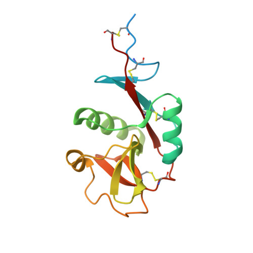
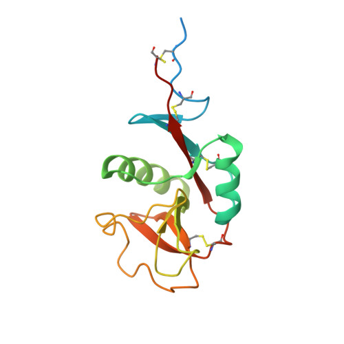
Structural Basis for Selective Recognition of Endogenous and Microbial Polysaccharides by Macrophage Receptor Sign-R1.

Silva-Martin, N.Bartual, S.G.Ramirez-Aportela, E.Chacon, P.Park, C.G.Hermoso, J.A.

(2014) Structure 22: 1595

  • DOI: https://doi.org/10.1016/j.str.2014.09.001
  • Primary Citation Related Structures: 
    3ZHG, 4C9F, 4CAJ, 4CDH

  • PubMed Abstract: 

    SIGN-R1 is a principal receptor for microbial polysaccharides uptake and is responsible for C3 fixation via an unusual complement activation pathway on splenic marginal zone macrophages. In these macrophages, SIGN-R1 is also involved in anti-inflammatory activity of intravenous immunoglobulin by direct interaction with sialylated Fcs. The high-resolution crystal structures of SIGN-R1 carbohydrate recognition domain and its complexes with dextran sulfate or sialic acid, and of the sialylated Fc antibody provide insights into SIGN-R1’s selective recognition of a-2,6-sialylated glycoproteins. Unexpectedly, an additional binding site has been found in the SIGNR1 carbohydrate recognition domain, structurally separate from the calcium-dependent carbohydrate-binding site. This secondary binding site could bind repetitive molecular patterns, as observed in microbial polysaccharides, in a calcium-independent manner. These two binding sites may allow SIGNR1 to simultaneously bind both immune glycoproteins and microbial polysaccharide components, accommodating SIGN-R1’s ability to relate the recognition of microbes to the activation of the classical complement pathway.

Macromolecule Content 

  • Total Structure Weight: 73.81 kDa 
  • Atom Count: 4,819 
  • Modeled Residue Count: 529 
  • Deposited Residue Count: 632 
  • Unique protein chains: 2

Macromolecules

Find similar proteins by:|  3D Structure
Entity ID: 1
MoleculeChains  Sequence LengthOrganismDetailsImage
CD209 ANTIGEN-LIKE PROTEIN B
A, B, C
158Mus musculusMutation(s): 0 
UniProt
Find proteins for Q8CJ91 (Mus musculus)
Explore Q8CJ91 
Go to UniProtKB:  Q8CJ91
Entity Groups
Sequence Clusters30% Identity50% Identity70% Identity90% Identity95% Identity100% Identity
UniProt GroupQ8CJ91
Sequence Annotations
Expand
Reference Sequence
Find similar proteins by:|  3D Structure
Entity ID: 2
MoleculeChains  Sequence LengthOrganismDetailsImage
CD209 ANTIGEN-LIKE PROTEIN B158Mus musculusMutation(s): 0 
UniProt
Find proteins for Q8CJ91 (Mus musculus)
Explore Q8CJ91 
Go to UniProtKB:  Q8CJ91
Entity Groups
Sequence Clusters30% Identity50% Identity70% Identity90% Identity95% Identity100% Identity
UniProt GroupQ8CJ91
Sequence Annotations
Expand
Reference Sequence

Small Molecules

Ligands 2 Unique
IDChains Name / Formula / InChI Key2D Diagram3D Interactions
SO4

Query on SO4


Download:Ideal Coordinates CCD File
E [auth A]
F [auth A]
G [auth A]
H [auth A]
I [auth A]
E [auth A],
F [auth A],
G [auth A],
H [auth A],
I [auth A],
J [auth A],
L [auth B],
M [auth B],
N [auth B],
P [auth C],
Q [auth C],
R [auth C],
S [auth C],
T [auth C],
U [auth C],
W [auth D],
X [auth D],
Y [auth D]
SULFATE ION
O4 S
QAOWNCQODCNURD-UHFFFAOYSA-L
CA

Query on CA


Download:Ideal Coordinates CCD File
K [auth A],
O [auth B],
V [auth C],
Z [auth D]
CALCIUM ION
Ca
BHPQYMZQTOCNFJ-UHFFFAOYSA-N

Experimental Data & Validation

Experimental Data

  • Method: X-RAY DIFFRACTION
  • Resolution: 1.87 Å
  • R-Value Free:  0.244 (Depositor), 0.240 (DCC) 
  • R-Value Work:  0.204 (Depositor), 0.201 (DCC) 
  • R-Value Observed: 0.207 (Depositor) 
Space Group: C 1 2 1
Unit Cell:
Length ( Å )Angle ( ˚ )
a = 146.72α = 90
b = 92.77β = 121.66
c = 77.06γ = 90
Software Package:
Software NamePurpose
PHENIXrefinement
MOSFLMdata reduction
SCALAdata scaling
MOLREPphasing

Structure Validation

View Full Validation Report


Entry History 

Deposition Data

Revision History  (Full details and data files)

  • Version 1.0: 2014-01-15
    Type: Initial release
  • Version 1.1: 2014-10-15
    Changes: Database references
  • Version 1.2: 2014-11-12
    Changes: Database references
  • Version 1.3: 2015-02-25
    Changes: Database references
  • Version 1.4: 2020-03-11
    Changes: Derived calculations, Other
  • Version 1.5: 2024-11-13
    Changes: Data collection, Database references, Derived calculations, Structure summary