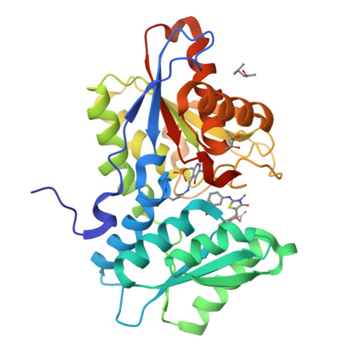
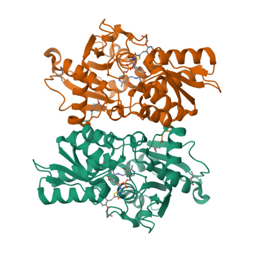
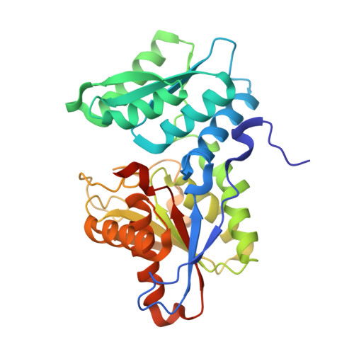
3ZEI

Structure of the Mycobacterium tuberculosis O-Acetylserine Sulfhydrylase (OASS) CysK1 in complex with a small molecule inhibitor


Experimental Data Snapshot

  • Method: X-RAY DIFFRACTION
  • Resolution: 2.00 Å
  • R-Value Free: 0.235 
  • R-Value Work: 0.202 
  • R-Value Observed: 0.203 

Starting Model: experimental
View more details

wwPDB Validation   3D Report Full Report


Ligand Structure Quality Assessment 


This is version 1.2 of the entry. See complete history


Literature

Structure-Guided Design of Novel Thiazolidine Inhibitors of O-Acetyl Serine Sulfhydrylase from Mycobacterium Tuberculosis.

Poyraz, O.Jeankumar, V.U.Saxena, S.Schnell, R.Haraldsson, M.Yogeeswari, P.Sriram, D.Schneider, G.

(2013) J Med Chem 56: 6457

  • DOI: https://doi.org/10.1021/jm400710k
  • Primary Citation of Related Structures:  
    3ZEI

  • PubMed Abstract: 

    The cysteine biosynthetic pathway is absent in humans but essential in microbial pathogens, suggesting that it provides potential targets for the development of novel antibacterial compounds. CysK1 is a pyridoxalphosphate-dependent O-acetyl sulfhydrylase, which catalyzes the formation of l-cysteine from O-acetyl serine and hydrogen sulfide. Here we report nanomolar thiazolidine inhibitors of Mycobacterium tuberculosis CysK1 developed by rational inhibitor design. The thiazolidine compounds were discovered using the crystal structure of a CysK1-peptide inhibitor complex as template. Pharmacophore modeling and subsequent in vitro screening resulted in an initial hit compound 2 (IC50 of 103.8 nM), which was subsequently optimized by a combination of protein crystallography, modeling, and synthetic chemistry. Hit expansion of 2 by chemical synthesis led to improved thiazolidine inhibitors with an IC50 value of 19 nM for the best compound, a 150-fold higher potency than the natural peptide inhibitor (IC50 2.9 μM).


  • Organizational Affiliation

    Division of Molecular Structural Biology, Department of Medical Biochemistry & Biophysics, Karolinska Institutet, S-171 77 Stockholm, Sweden.


Macromolecules
Find similar proteins by:  (by identity cutoff)  |  3D Structure
Entity ID: 1
MoleculeChains Sequence LengthOrganismDetailsImage
O-ACETYLSERINE SULFHYDRYLASE310Mycobacterium tuberculosisMutation(s): 0 
EC: 2.5.1.47
UniProt
Find proteins for P9WP55 (Mycobacterium tuberculosis (strain ATCC 25618 / H37Rv))
Explore P9WP55 
Go to UniProtKB:  P9WP55
Entity Groups  
Sequence Clusters30% Identity50% Identity70% Identity90% Identity95% Identity100% Identity
UniProt GroupP9WP55
Sequence Annotations
Expand
  • Reference Sequence
Small Molecules
Binding Affinity Annotations 
IDSourceBinding Affinity
AWH BindingDB:  3ZEI IC50: 103 (nM) from 1 assay(s)
Experimental Data & Validation

Experimental Data

  • Method: X-RAY DIFFRACTION
  • Resolution: 2.00 Å
  • R-Value Free: 0.235 
  • R-Value Work: 0.202 
  • R-Value Observed: 0.203 
  • Space Group: P 41 21 2
Unit Cell:
Length ( Å )Angle ( ˚ )
a = 71.82α = 90
b = 71.82β = 90
c = 179.63γ = 90
Software Package:
Software NamePurpose
REFMACrefinement
MOSFLMdata reduction
SCALEPACKdata scaling
PHASERphasing

Structure Validation

View Full Validation Report


Ligand Structure Quality Assessment 


Entry History 

Deposition Data

Revision History  (Full details and data files)

  • Version 1.0: 2013-08-07
    Type: Initial release
  • Version 1.1: 2013-09-04
    Changes: Database references
  • Version 1.2: 2023-12-20
    Changes: Data collection, Database references, Derived calculations, Other, Refinement description