3X24 | pdb_00003x24

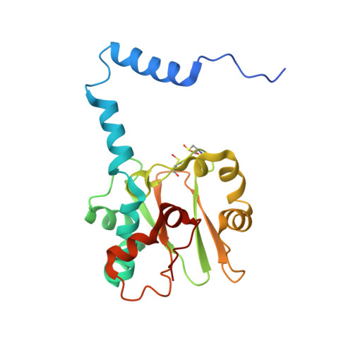
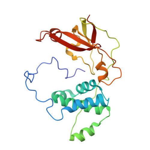
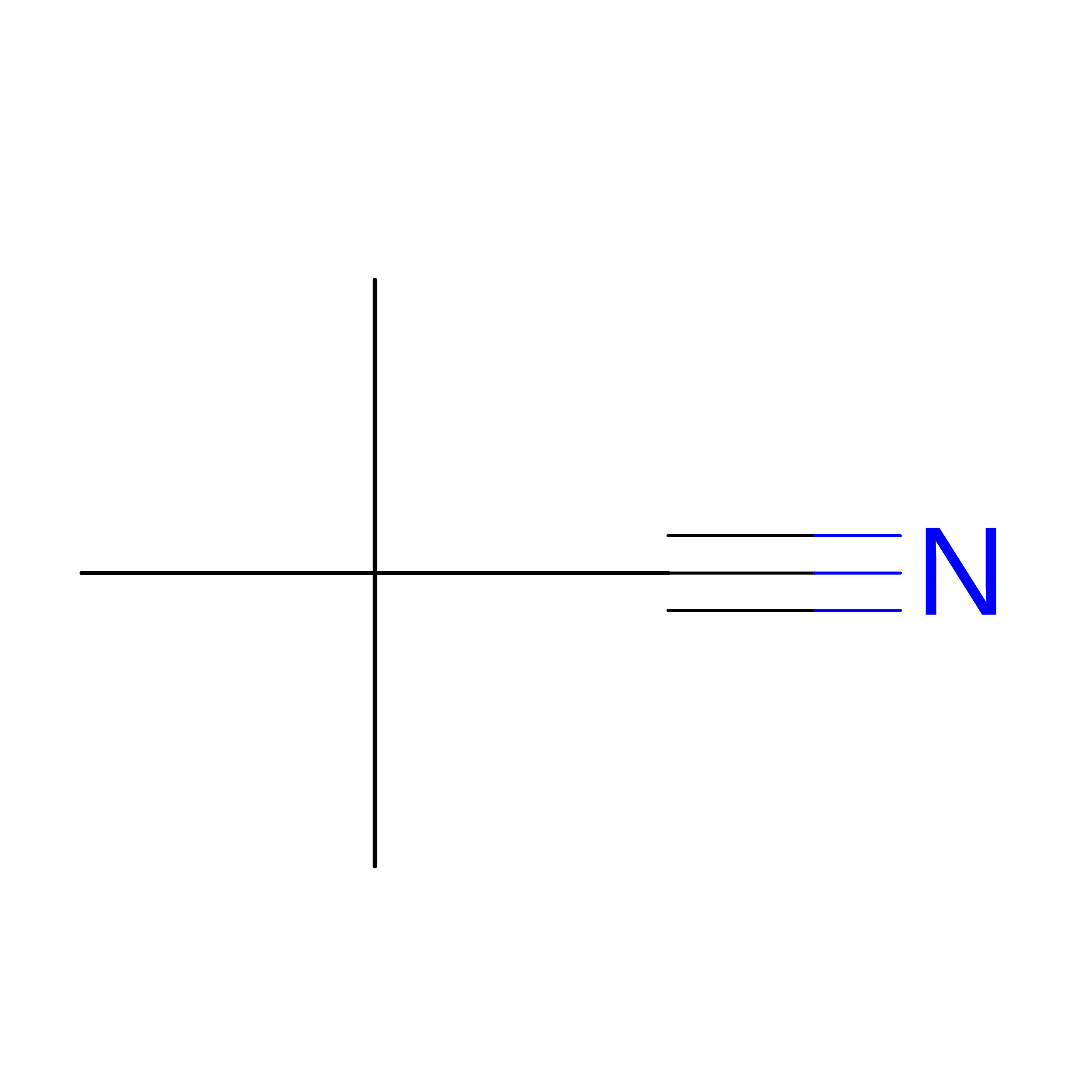
Crystal structure of Nitrile Hydratase mutant bR56K complexed with Trimethylacetonitrile, photo-activated for 120 min


Experimental Data Snapshot

  • Method: X-RAY DIFFRACTION
  • Resolution: 1.24 Å
  • R-Value Free: 
    0.163 (Depositor), 0.169 (DCC) 
  • R-Value Work: 
    0.138 (Depositor), 0.144 (DCC) 
  • R-Value Observed: 
    0.139 (Depositor) 

wwPDB Validation 3D Report Full Report

Validation slider image for 3X24

This is version 1.1 of the entry. See complete history. 

Literature

Time-Resolved Crystallography of the Reaction Intermediate of Nitrile Hydratase: Revealing a Role for the Cysteinesulfenic Acid Ligand as a Catalytic Nucleophile.

Yamanaka, Y., Kato, Y., Hashimoto, K., Iida, K., Nagasawa, K., Nakayama, H., Dohmae, N., Noguchi, K., Noguchi, T., Yohda, M., Odaka, M.

(2015) Angew Chem Int Ed Engl 54: 10763-10767

  • DOI: https://doi.org/10.1002/anie.201502731
  • Primary Citation Related Structures: 
    3WVD, 3WVE, 3X20, 3X24, 3X25, 3X26

  • PubMed Abstract: 

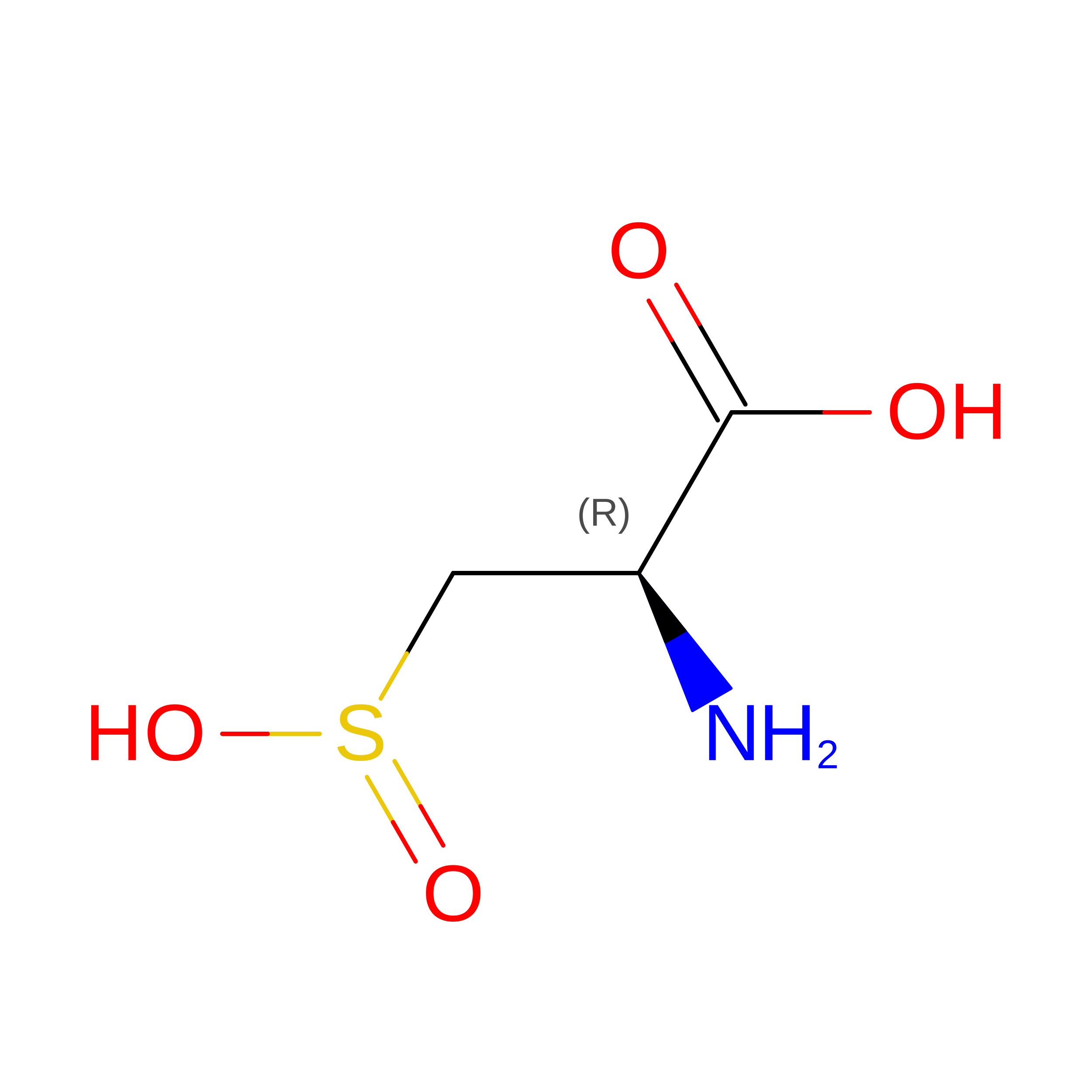
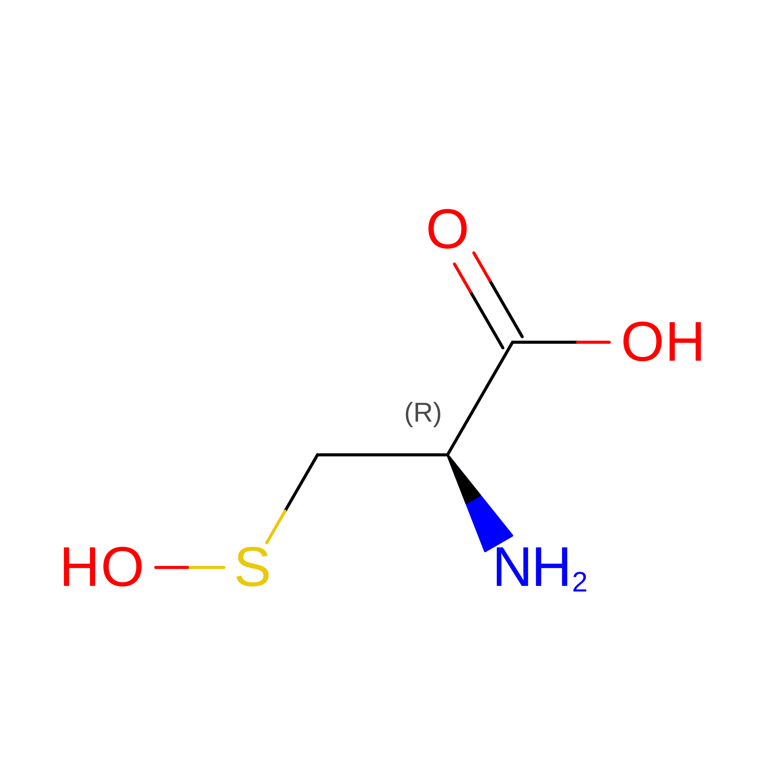
    The reaction mechanism of nitrile hydratase (NHase) was investigated using time-resolved crystallography of the mutant NHase, in which βArg56, strictly conserved and hydrogen bonded to the two post-translationally oxidized cysteine ligands, was replaced by lysine, and pivalonitrile was the substrate. The crystal structures of the reaction intermediates were determined at high resolution (1.2-1.3 Å). In combination with FTIR analyses of NHase following hydration in H2 (18) O, we propose that the metal-coordinated substrate is nucleophilically attacked by the O(SO(-) ) atom of αCys114-SO(-) , followed by nucleophilic attack of the S(SO(-) ) atom by a βArg56-activated water molecule to release the product amide and regenerate αCys114-SO(-) .


  • Organizational Affiliation: 
    • Department of Biotechnology and Life Science, Graduate School of Technology, Tokyo University of Agriculture and Technology, Koganei, Tokyo 184-8588 (Japan).

Macromolecule Content 

  • Total Structure Weight: 46.74 kDa 
  • Atom Count: 3,676 
  • Modeled Residue Count: 409 
  • Deposited Residue Count: 419 
  • Unique protein chains: 2

Macromolecules

Find similar proteins by:|  3D Structure
Entity ID: 1
MoleculeChains  Sequence LengthOrganismDetailsImage
Nitrile hydratase subunit alpha207Rhodococcus erythropolisMutation(s): 0 
Gene Names: nthA
EC: 4.2.1.84
UniProt
Find proteins for P13448 (Rhodococcus erythropolis)
Explore P13448 
Go to UniProtKB:  P13448
Entity Groups
Sequence Clusters30% Identity50% Identity70% Identity90% Identity95% Identity100% Identity
UniProt GroupP13448
Sequence Annotations
Expand
Reference Sequence
Find similar proteins by:|  3D Structure
Entity ID: 2
MoleculeChains  Sequence LengthOrganismDetailsImage
Nitrile hydratase subunit beta212Rhodococcus erythropolisMutation(s): 1 
Gene Names: nthB, nha2
EC: 4.2.1.84
UniProt
Find proteins for P13449 (Rhodococcus erythropolis)
Explore P13449 
Go to UniProtKB:  P13449
Entity Groups
Sequence Clusters30% Identity50% Identity70% Identity90% Identity95% Identity100% Identity
UniProt GroupP13449
Sequence Annotations
Expand
Reference Sequence

Experimental Data & Validation

Experimental Data

  • Method: X-RAY DIFFRACTION
  • Resolution: 1.24 Å
  • R-Value Free:  0.163 (Depositor), 0.169 (DCC) 
  • R-Value Work:  0.138 (Depositor), 0.144 (DCC) 
  • R-Value Observed: 0.139 (Depositor) 
Space Group: C 1 2 1
Unit Cell:
Length ( Å )Angle ( ˚ )
a = 113.91α = 90
b = 59.913β = 125.05
c = 81.585γ = 90
Software Package:
Software NamePurpose
HKL-2000data collection
MOLREPphasing
REFMACrefinement
HKL-2000data reduction
HKL-2000data scaling

Structure Validation

View Full Validation Report


Entry History 

Deposition Data

Revision History  (Full details and data files)

  • Version 1.0: 2016-01-27
    Type: Initial release
  • Version 1.1: 2024-10-16
    Changes: Data collection, Database references, Derived calculations, Structure summary