3WZU | pdb_00003wzu

THE STRUCTURE OF MAP2K7 IN COMPLEX WITH 5Z-7-oxozeaenol


Experimental Data Snapshot

  • Method: X-RAY DIFFRACTION
  • Resolution: 3.01 Å
  • R-Value Free: 
    0.322 (Depositor), 0.322 (DCC) 
  • R-Value Work: 
    0.236 (Depositor), 0.232 (DCC) 
  • R-Value Observed: 
    0.240 (Depositor) 

Starting Model: experimental
View more details

wwPDB Validation 3D Report Full Report

Validation slider image for 3WZU

Ligand Structure Quality Assessment 


This is version 1.3 of the entry. See complete history

Literature

5Z-7-Oxozeaenol covalently binds to MAP2K7 at Cys218 in an unprecedented manner.

Sogabe, Y.Matsumoto, T.Hashimoto, T.Kirii, Y.Sawa, M.Kinoshita, T.

(2015) Bioorg Med Chem Lett 25: 593-596

  • DOI: https://doi.org/10.1016/j.bmcl.2014.12.011
  • Primary Citation Related Structures: 
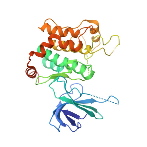
    3WZU

  • PubMed Abstract: 

    5Z-7-Oxozeaenol (5Z7O) is a covalent bonding inhibitor against the several protein kinases (e.g., ERK2 and TAK1) that possess a free cysteine at the gatekeeper-2 position. In addition to this cysteine, MAP2K7 has three other cysteine residues that are candidate for covalent bonding by the inhibitor 5Z7O. The crystal structure of the MAP2K7/5Z7O complex revealed that the inhibitor binds to MAP2K7 at a cysteine residue located at the end of the hinge region and not at the gatekeeper-2 residue. The structural insights into the interaction of 5Z7O with MAP2K7 should aid the development of 5Z7O derivatives with improved potency and selectivity.


  • Organizational Affiliation
    • Graduate School of Science, Osaka Prefecture University, 1-1 Gakuen-cho, Naka-ku, Sakai, Osaka 599-8531, Japan.

Macromolecule Content 

  • Total Structure Weight: 37.36 kDa 
  • Atom Count: 2,200 
  • Modeled Residue Count: 270 
  • Deposited Residue Count: 324 
  • Unique protein chains: 1

Macromolecules

Find similar proteins by:|  3D Structure
Entity ID: 1
MoleculeChains  Sequence LengthOrganismDetailsImage
Dual specificity mitogen-activated protein kinase kinase 7324Homo sapiensMutation(s): 0 
Gene Names: MAP2K7JNKK2MEK7MKK7PRKMK7SKK4
EC: 2.7.12.2
UniProt & NIH Common Fund Data Resources
Find proteins for O14733 (Homo sapiens)
Explore O14733 
Go to UniProtKB:  O14733
PHAROS:  O14733
GTEx:  ENSG00000076984 
Entity Groups
Sequence Clusters30% Identity50% Identity70% Identity90% Identity95% Identity100% Identity
UniProt GroupO14733
Sequence Annotations
Expand
Reference Sequence

Small Molecules

Ligands 1 Unique
IDChains Name / Formula / InChI Key2D Diagram3D Interactions
1FM

Query on 1FM


Download:Ideal Coordinates CCD File
B [auth A](3S,5Z,8S,9S,11E)-8,9,16-trihydroxy-14-methoxy-3-methyl-3,4,9,10-tetrahydro-1H-2-benzoxacyclotetradecine-1,7(8H)-dione
C19 H22 O7
NEQZWEXWOFPKOT-BYRRXHGESA-N
Binding Affinity Annotations 
IDSourceBinding Affinity
1FM BindingDB:  3WZU IC50: 1300 (nM) from 1 assay(s)

Experimental Data & Validation

Experimental Data

  • Method: X-RAY DIFFRACTION
  • Resolution: 3.01 Å
  • R-Value Free:  0.322 (Depositor), 0.322 (DCC) 
  • R-Value Work:  0.236 (Depositor), 0.232 (DCC) 
  • R-Value Observed: 0.240 (Depositor) 
Space Group: P 61 2 2
Unit Cell:
Length ( Å )Angle ( ˚ )
a = 71.395α = 90
b = 71.395β = 90
c = 268.817γ = 120
Software Package:
Software NamePurpose
ADSCdata collection
MOLREPphasing
REFMACrefinement
HKL-2000data reduction
SCALEPACKdata scaling

Structure Validation

View Full Validation Report


Ligand Structure Quality Assessment 


Entry History 

Deposition Data

Revision History  (Full details and data files)

  • Version 1.0: 2015-01-14
    Type: Initial release
  • Version 1.1: 2017-11-22
    Changes: Refinement description
  • Version 1.2: 2022-08-24
    Changes: Database references, Derived calculations
  • Version 1.3: 2023-11-08
    Changes: Data collection, Refinement description