3WZT | pdb_00003wzt

Crystal structure of Trx3 domain of UGGT (detergent-unbound form)


Experimental Data Snapshot

  • Method: X-RAY DIFFRACTION
  • Resolution: 3.40 Å
  • R-Value Free: 
    0.292 (Depositor), 0.291 (DCC) 
  • R-Value Work: 
    0.235 (Depositor), 0.234 (DCC) 
  • R-Value Observed: 
    0.237 (Depositor) 

Starting Model: experimental
View more details

wwPDB Validation 3D Report Full Report

Validation slider image for 3WZT

This is version 1.4 of the entry. See complete history

Literature

Structural insight into substrate recognition by the endoplasmic reticulum folding-sensor enzyme: crystal structure of third thioredoxin-like domain of UDP-glucose:glycoprotein glucosyltransferase

Zhu, T.Satoh, T.Kato, K.

(2014) Sci Rep 4: 7322-7322

  • DOI: https://doi.org/10.1038/srep07322
  • Primary Citation Related Structures: 
    3WZS, 3WZT

  • PubMed Abstract: 

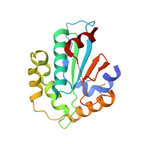
    The endoplasmic reticulum (ER) possesses a protein quality control system that supports the efficient folding of newly synthesized glycoproteins. In this system, a series of N-linked glycan intermediates displayed on proteins serve as quality tags. The ER folding-sensor enzyme UDP-glucose:glycoprotein glucosyltransferase (UGGT) operates as the gatekeeper for ER quality control by specifically transferring monoglucose residues to incompletely folded glycoproteins, thereby allowing them to interact with lectin chaperone complexes to facilitate their folding. Despite its functional importance, no structural information is available for this key enzyme to date. To elucidate the folding-sensor mechanism in the ER, we performed a structural study of UGGT. Based on bioinformatics analyses, the folding-sensor region of UGGT was predicted to harbour three tandem thioredoxin (Trx)-like domains, which are often found in proteins involved in ER quality control. Furthermore, we determined the three-dimensional structure of the third Trx-like domain, which exhibits an extensive hydrophobic patch concealed by its flexible C-terminal helix. Our structural data suggest that this hydrophobic patch is involved in intermolecular interactions, thereby contributing to the folding-sensor mechanism of UGGT.


  • Organizational Affiliation
    • 1] School of Physical Sciences, The Graduate University for Advanced Studies, 5-1 Higashiyama, Myodaiji, Okazaki, Aichi 444-8787, Japan [2] Okazaki Institute for Integrative Bioscience and Institute for Molecular Science, National Institutes of Natural Sciences, 5-1 Higashiyama, Myodaiji, Okazaki, Aichi 444-8787, Japan [3] Graduate School of Pharmaceutical Sciences, Nagoya City University, 3-1 Tanabe-dori, Mizuho-ku, Nagoya 467-8603, Japan.

Macromolecule Content 

  • Total Structure Weight: 110.34 kDa 
  • Atom Count: 6,452 
  • Modeled Residue Count: 812 
  • Deposited Residue Count: 978 
  • Unique protein chains: 1

Macromolecules

Find similar proteins by:|  3D Structure
Entity ID: 1
MoleculeChains  Sequence LengthOrganismDetailsImage
UDP-glucose-glycoprotein glucosyltransferase-like protein
A, B, C, D, E
A, B, C, D, E, F
163Thermochaetoides thermophila DSM 1495Mutation(s): 0 
Gene Names: CTHT_0048990
UniProt
Find proteins for G0SB58 (Chaetomium thermophilum (strain DSM 1495 / CBS 144.50 / IMI 039719))
Explore G0SB58 
Go to UniProtKB:  G0SB58
Entity Groups
Sequence Clusters30% Identity50% Identity70% Identity90% Identity95% Identity100% Identity
UniProt GroupG0SB58
Sequence Annotations
Expand
Reference Sequence

Small Molecules

Modified Residues  1 Unique
IDChains TypeFormula2D DiagramParent
MSE
Query on MSE
A, B, C, D, E
A, B, C, D, E, F
L-PEPTIDE LINKINGC5 H11 N O2 SeMET

Experimental Data & Validation

Experimental Data

  • Method: X-RAY DIFFRACTION
  • Resolution: 3.40 Å
  • R-Value Free:  0.292 (Depositor), 0.291 (DCC) 
  • R-Value Work:  0.235 (Depositor), 0.234 (DCC) 
  • R-Value Observed: 0.237 (Depositor) 
Space Group: I 2 3
Unit Cell:
Length ( Å )Angle ( ˚ )
a = 196.4α = 90
b = 196.4β = 90
c = 196.4γ = 90
Software Package:
Software NamePurpose
HKL-2000data collection
PHASERphasing
REFMACrefinement
DENZOdata reduction
SCALEPACKdata scaling

Structure Validation

View Full Validation Report


Entry History 

Deposition Data

Revision History  (Full details and data files)

  • Version 1.0: 2014-12-03
    Type: Initial release
  • Version 1.1: 2014-12-31
    Changes: Database references
  • Version 1.2: 2023-11-08
    Changes: Data collection, Database references, Derived calculations, Refinement description
  • Version 1.3: 2023-12-06
    Changes: Data collection
  • Version 1.4: 2024-11-20
    Changes: Structure summary