3WZ1 | pdb_00003wz1

Catalytic domain of beta-agarase from Microbulbifer thermotolerans JAMB-A94


Experimental Data Snapshot

  • Method: X-RAY DIFFRACTION
  • Resolution: 1.60 Å
  • R-Value Free: 
    0.165 (Depositor), 0.173 (DCC) 
  • R-Value Work: 
    0.152 (Depositor), 0.165 (DCC) 
  • R-Value Observed: 
    0.153 (Depositor) 

Starting Model: experimental
View more details

wwPDB Validation 3D Report Full Report

Validation slider image for 3WZ1

This is version 1.2 of the entry. See complete history

Literature

Crystal structure of the catalytic domain of a GH16 beta-agarase from a deep-sea bacterium, Microbulbifer thermotolerans JAMB-A94

Takagi, E.Hatada, Y.Akita, M.Ohta, Y.Yokoi, G.Miyazaki, T.Nishikawa, A.Tonozuka, T.

(2015) Biosci Biotechnol Biochem 79: 625-632

  • DOI: https://doi.org/10.1080/09168451.2014.988680
  • Primary Citation Related Structures: 
    3WZ1

  • PubMed Abstract: 

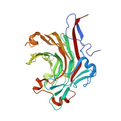
    A deep-sea bacterium, Microbulbifer thermotolerans JAMB-A94, has a β-agarase (MtAgaA) belonging to the glycoside hydrolase family (GH) 16. The optimal temperature of this bacterium for growth is 43-49 °C, and MtAgaA is stable at 60 °C, which is one of the most thermostable enzymes among GH16 β-agarases. Here, we determined the catalytic domain structure of MtAgaA. MtAgaA consists of a β-jelly roll fold, as observed in other GH16 enzymes. The structure of MtAgaA was most similar to two β-agarases from Zobellia galactanivorans, ZgAgaA, and ZgAgaB. Although the catalytic cleft structure of MtAgaA was similar to ZgAgaA and ZgAgaB, residues at subsite -4 of MtAgaA were not conserved between them. Also, an α-helix, designated as α4', was uniquely located near the catalytic cleft of MtAgaA. A comparison of the structures of the three enzymes suggested that multiple factors, including increased numbers of arginine and proline residues, could contribute to the thermostability of MtAgaA.


  • Organizational Affiliation
    • a Japan Agency for Marine-Earth Science and Technology (JAMSTEC) , Yokosuka , Japan.

Macromolecule Content 

  • Total Structure Weight: 32.26 kDa 
  • Atom Count: 2,533 
  • Modeled Residue Count: 277 
  • Deposited Residue Count: 282 
  • Unique protein chains: 1

Macromolecules

Find similar proteins by:|  3D Structure
Entity ID: 1
MoleculeChains  Sequence LengthOrganismDetailsImage
Agarase282Microbulbifer thermotoleransMutation(s): 0 
Gene Names: agaA
EC: 3.2.1.81
UniProt
Find proteins for Q6F4N4 (Microbulbifer thermotolerans)
Explore Q6F4N4 
Go to UniProtKB:  Q6F4N4
Entity Groups
Sequence Clusters30% Identity50% Identity70% Identity90% Identity95% Identity100% Identity
UniProt GroupQ6F4N4
Sequence Annotations
Expand
Reference Sequence

Experimental Data & Validation

Experimental Data

  • Method: X-RAY DIFFRACTION
  • Resolution: 1.60 Å
  • R-Value Free:  0.165 (Depositor), 0.173 (DCC) 
  • R-Value Work:  0.152 (Depositor), 0.165 (DCC) 
  • R-Value Observed: 0.153 (Depositor) 
Space Group: P 21 21 21
Unit Cell:
Length ( Å )Angle ( ˚ )
a = 49.624α = 90
b = 69.394β = 90
c = 90.367γ = 90
Software Package:
Software NamePurpose
ADSCdata collection
MOLREPphasing
REFMACrefinement
HKL-2000data reduction
HKL-2000data scaling

Structure Validation

View Full Validation Report


Entry History 

Deposition Data

Revision History  (Full details and data files)

  • Version 1.0: 2014-11-19
    Type: Initial release
  • Version 1.1: 2015-04-22
    Changes: Database references
  • Version 1.2: 2023-11-08
    Changes: Data collection, Database references, Derived calculations, Refinement description