3WYW | pdb_00003wyw

Structural characterization of catalytic site of a Nilaparvata lugens delta-class glutathione transferase


Experimental Data Snapshot

  • Method: X-RAY DIFFRACTION
  • Resolution: 1.70 Å
  • R-Value Free: 
    0.217 (Depositor), 0.231 (DCC) 
  • R-Value Work: 
    0.175 (Depositor) 
  • R-Value Observed: 
    0.177 (Depositor) 

wwPDB Validation 3D Report Full Report

Validation slider image for 3WYW

Ligand Structure Quality Assessment 


This is version 1.1 of the entry. See complete history

Literature

Structural characterization of the catalytic site of a Nilaparvata lugens delta-class glutathione transferase.

Yamamoto, K.Higashiura, A.Hossain, M.T.Yamada, N.Shiotsuki, T.Nakagawa, A.

(2014) Arch Biochem Biophys 566C: 36-42

  • DOI: https://doi.org/10.1016/j.abb.2014.12.001
  • Primary Citation Related Structures: 
    3WYW

  • PubMed Abstract: 

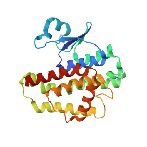
    Glutathione transferases (GSTs) are a major class of detoxification enzymes that play a central role in the defense against environmental toxicants and oxidative stress. Here, we studied the crystal structure of a delta-class glutathione transferase from Nilaparvata lugens, nlGSTD, to gain insights into its catalytic mechanism. The structure of nlGSTD in complex with glutathione, determined at a resolution of 1.7Å, revealed that it exists as a dimer and its secondary and tertiary structures are similar to those of other delta-class GSTs. Analysis of a complex between nlGSTD and glutathione showed that the bound glutathione was localized to the glutathione-binding site. Site-directed mutagenesis of nlGSTD mutants indicated that amino acid residues Ser11, His52, Glu66, and Phe119 contribute to catalytic activity.


  • Organizational Affiliation
    • Faculty of Agriculture, Kyushu University Graduate School, 6-10-1 Hakozaki, Higashi-ku, Fukuoka 812-8581, Japan. Electronic address: yamamok@agr.kyushu-u.ac.jp.

Macromolecule Content 

  • Total Structure Weight: 48.62 kDa 
  • Atom Count: 3,795 
  • Modeled Residue Count: 427 
  • Deposited Residue Count: 432 
  • Unique protein chains: 1

Macromolecules

Find similar proteins by:|  3D Structure
Entity ID: 1
MoleculeChains  Sequence LengthOrganismDetailsImage
Glutathione S-transferase
A, B
216Nilaparvata lugensMutation(s): 0 
UniProt
Find proteins for Q8N0B3 (Nilaparvata lugens)
Explore Q8N0B3 
Go to UniProtKB:  Q8N0B3
Entity Groups
Sequence Clusters30% Identity50% Identity70% Identity90% Identity95% Identity100% Identity
UniProt GroupQ8N0B3
Sequence Annotations
Expand
Reference Sequence

Biologically Interesting Molecules (External Reference) 

1 Unique

Experimental Data & Validation

Experimental Data

  • Method: X-RAY DIFFRACTION
  • Resolution: 1.70 Å
  • R-Value Free:  0.217 (Depositor), 0.231 (DCC) 
  • R-Value Work:  0.175 (Depositor) 
  • R-Value Observed: 0.177 (Depositor) 
Space Group: P 1 21 1
Unit Cell:
Length ( Å )Angle ( ˚ )
a = 54.552α = 90
b = 49.104β = 99.9
c = 74.184γ = 90
Software Package:
Software NamePurpose
HKL-2000data collection
MERLOTphasing
REFMACrefinement
HKL-2000data reduction
HKL-2000data scaling

Structure Validation

View Full Validation Report


Ligand Structure Quality Assessment 


Entry History 

Deposition Data

Revision History  (Full details and data files)

  • Version 1.0: 2015-01-14
    Type: Initial release
  • Version 1.1: 2024-03-20
    Changes: Data collection, Database references, Derived calculations