3WX2 | pdb_00003wx2

Mouse Cereblon thalidomide binding domain, native


Experimental Data Snapshot

  • Method: X-RAY DIFFRACTION
  • Resolution: 2.00 Å
  • R-Value Free: 
    0.225 (Depositor), 0.225 (DCC) 
  • R-Value Work: 
    0.195 (Depositor), 0.196 (DCC) 
  • R-Value Observed: 
    0.197 (Depositor) 

Starting Model: experimental
View more details

wwPDB Validation 3D Report Full Report

Validation slider image for 3WX2

This is version 1.3 of the entry. See complete history

Literature

Structure of the human Cereblon-DDB1-lenalidomide complex reveals basis for responsiveness to thalidomide analogs

Chamberlain, P.P.Lopez-Girona, A.Miller, K.Carmel, G.Pagarigan, B.Chie-Leon, B.Rychak, E.Corral, L.G.Ren, Y.J.Wang, M.Riley, M.Delker, S.L.Ito, T.Ando, H.Mori, T.Hirano, Y.Handa, H.Hakoshima, T.Daniel, T.O.Cathers, B.E.

(2014) Nat Struct Mol Biol 21: 803-809

  • DOI: https://doi.org/10.1038/nsmb.2874
  • Primary Citation Related Structures: 
    3WX1, 3WX2

  • PubMed Abstract: 

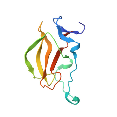
    The Cul4-Rbx1-DDB1-Cereblon E3 ubiquitin ligase complex is the target of thalidomide, lenalidomide and pomalidomide, therapeutically important drugs for multiple myeloma and other B-cell malignancies. These drugs directly bind Cereblon (CRBN) and promote the recruitment of substrates Ikaros (IKZF1) and Aiolos (IKZF3) to the E3 complex, thus leading to substrate ubiquitination and degradation. Here we present the crystal structure of human CRBN bound to DDB1 and the drug lenalidomide. A hydrophobic pocket in the thalidomide-binding domain (TBD) of CRBN accommodates the glutarimide moiety of lenalidomide, whereas the isoindolinone ring is exposed to solvent. We also solved the structures of the mouse TBD in the apo state and with thalidomide or pomalidomide. Site-directed mutagenesis in lentiviral-expression myeloma models showed that key drug-binding residues are critical for antiproliferative effects.


  • Organizational Affiliation
    • Celgene Corporation, San Diego, California, USA.

Macromolecule Content 

  • Total Structure Weight: 25.19 kDa 
  • Atom Count: 1,773 
  • Modeled Residue Count: 207 
  • Deposited Residue Count: 222 
  • Unique protein chains: 1

Macromolecules

Find similar proteins by:|  3D Structure
Entity ID: 1
MoleculeChains  Sequence LengthOrganismDetailsImage
Protein cereblon
A, B
111Mus musculusMutation(s): 0 
Gene Names: Crbn
UniProt & NIH Common Fund Data Resources
Find proteins for Q8C7D2 (Mus musculus)
Explore Q8C7D2 
Go to UniProtKB:  Q8C7D2
IMPC:  MGI:1913277
Entity Groups
Sequence Clusters30% Identity50% Identity70% Identity90% Identity95% Identity100% Identity
UniProt GroupQ8C7D2
Sequence Annotations
Expand
Reference Sequence

Experimental Data & Validation

Experimental Data

  • Method: X-RAY DIFFRACTION
  • Resolution: 2.00 Å
  • R-Value Free:  0.225 (Depositor), 0.225 (DCC) 
  • R-Value Work:  0.195 (Depositor), 0.196 (DCC) 
  • R-Value Observed: 0.197 (Depositor) 
Space Group: I 21 3
Unit Cell:
Length ( Å )Angle ( ˚ )
a = 117.726α = 90
b = 117.726β = 90
c = 117.726γ = 90
Software Package:
Software NamePurpose
HKL-2000data collection
PHASERphasing
REFMACrefinement
HKL-2000data reduction
HKL-2000data scaling

Structure Validation

View Full Validation Report


Entry History 

Deposition Data

Revision History  (Full details and data files)

  • Version 1.0: 2014-08-06
    Type: Initial release
  • Version 1.1: 2014-08-27
    Changes: Database references
  • Version 1.2: 2014-09-17
    Changes: Database references
  • Version 1.3: 2023-11-08
    Changes: Data collection, Database references, Derived calculations, Refinement description