3WV5 | pdb_00003wv5

Complex structure of VinN with 3-methylaspartate


Experimental Data Snapshot

  • Method: X-RAY DIFFRACTION
  • Resolution: 2.20 Å
  • R-Value Free: 
    0.245 (Depositor), 0.242 (DCC) 
  • R-Value Work: 
    0.205 (Depositor) 
  • R-Value Observed: 
    0.207 (Depositor) 

Starting Model: experimental
View more details

wwPDB Validation 3D Report Full Report

Validation slider image for 3WV5

This is version 1.2 of the entry. See complete history

Literature

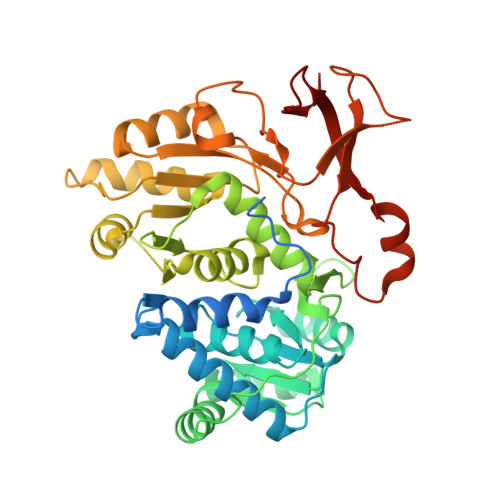
The crystal structure of the adenylation enzyme VinN reveals a unique beta-amino acid recognition mechanism

Miyanaga, A.Cieslak, J.Shinohara, Y.Kudo, F.Eguchi, T.

(2014) J Biological Chem 289: 31448-31457

  • DOI: https://doi.org/10.1074/jbc.M114.602326
  • Primary Citation Related Structures: 
    3WV4, 3WV5, 3WVN

  • PubMed Abstract: 

    Adenylation enzymes play important roles in the biosynthesis and degradation of primary and secondary metabolites. Mechanistic insights into the recognition of α-amino acid substrates have been obtained for α-amino acid adenylation enzymes. The Asp residue is invariant and is essential for the stabilization of the α-amino group of the substrate. In contrast, the β-amino acid recognition mechanism of adenylation enzymes is still unclear despite the importance of β-amino acid activation for the biosynthesis of various natural products. Herein, we report the crystal structure of the stand-alone adenylation enzyme VinN, which specifically activates (2S,3S)-3-methylaspartate (3-MeAsp) in vicenistatin biosynthesis. VinN has an overall structure similar to that of other adenylation enzymes. The structure of the complex with 3-MeAsp revealed that a conserved Asp(230) residue is used in the recognition of the β-amino group of 3-MeAsp similar to α-amino acid adenylation enzymes. A mutational analysis and structural comparison with α-amino acid adenylation enzymes showed that the substrate-binding pocket of VinN has a unique architecture to accommodate 3-MeAsp as a β-amino acid substrate. Thus, the VinN structure allows the first visualization of the interaction of an adenylation enzyme with a β-amino acid and provides new mechanistic insights into the selective recognition of β-amino acids in this family of enzymes.


  • Organizational Affiliation
    • From the Departments of Chemistry and.

Macromolecule Content 

  • Total Structure Weight: 96.64 kDa 
  • Atom Count: 5,913 
  • Modeled Residue Count: 746 
  • Deposited Residue Count: 884 
  • Unique protein chains: 1

Macromolecules

Find similar proteins by:|  3D Structure
Entity ID: 1
MoleculeChains  Sequence LengthOrganismDetailsImage
Non-ribosomal peptide synthetase
A, B
442Streptomyces halstediiMutation(s): 0 
Gene Names: vinN
EC: 6.2.1
UniProt
Find proteins for Q76KY2 (Streptomyces halstedii)
Explore Q76KY2 
Go to UniProtKB:  Q76KY2
Entity Groups
Sequence Clusters30% Identity50% Identity70% Identity90% Identity95% Identity100% Identity
UniProt GroupQ76KY2
Sequence Annotations
Expand
Reference Sequence

Experimental Data & Validation

Experimental Data

  • Method: X-RAY DIFFRACTION
  • Resolution: 2.20 Å
  • R-Value Free:  0.245 (Depositor), 0.242 (DCC) 
  • R-Value Work:  0.205 (Depositor) 
  • R-Value Observed: 0.207 (Depositor) 
Space Group: C 2 2 21
Unit Cell:
Length ( Å )Angle ( ˚ )
a = 80.66α = 90
b = 109.41β = 90
c = 200.21γ = 90
Software Package:
Software NamePurpose
ADSCdata collection
MOLREPphasing
REFMACrefinement
MOSFLMdata reduction
SCALAdata scaling

Structure Validation

View Full Validation Report


Entry History 

Deposition Data

Revision History  (Full details and data files)

  • Version 1.0: 2014-10-01
    Type: Initial release
  • Version 1.1: 2015-02-11
    Changes: Database references
  • Version 1.2: 2023-11-08
    Changes: Data collection, Database references, Derived calculations, Refinement description