3WSY

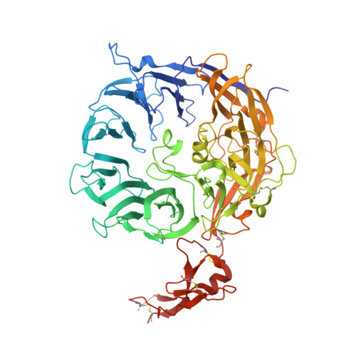
SorLA Vps10p domain in complex with its own propeptide fragment


Experimental Data Snapshot

  • Method: X-RAY DIFFRACTION
  • Resolution: 3.11 Å
  • R-Value Free: 0.232 
  • R-Value Work: 0.195 
  • R-Value Observed: 0.197 

Starting Model: experimental
View more details

wwPDB Validation   3D Report Full Report


Ligand Structure Quality Assessment 


This is version 1.4 of the entry. See complete history


Literature

Structural basis for amyloidogenic peptide recognition by sorLA.

Kitago, Y.Nagae, M.Nakata, Z.Yagi-Utsumi, M.Takagi-Niidome, S.Mihara, E.Nogi, T.Kato, K.Takagi, J.

(2015) Nat Struct Mol Biol 22: 199-206

  • DOI: https://doi.org/10.1038/nsmb.2954
  • Primary Citation of Related Structures:  
    3WSX, 3WSY, 3WSZ

  • PubMed Abstract: 

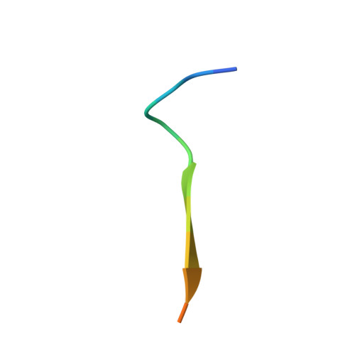
    SorLA is a neuronal sorting receptor considered to be a major risk factor for Alzheimer's disease. We have recently reported that it directs lysosomal targeting of nascent neurotoxic amyloid-β (Aβ) peptides by directly binding Aβ. Here, we determined the crystal structure of the human sorLA domain responsible for Aβ capture, Vps10p, in an unbound state and in complex with two ligands. Vps10p assumes a ten-bladed β-propeller fold with a large tunnel at the center. An internal ligand derived from the sorLA propeptide bound inside the tunnel to extend the β-sheet of one of the propeller blades. The structure of the sorLA Vps10p-Aβ complex revealed that the same site is used. Peptides are recognized by sorLA Vps10p in redundant modes without strict dependence on a particular amino acid sequence, thus suggesting a broad specificity toward peptides with a propensity for β-sheet formation.


  • Organizational Affiliation

    Institute for Protein Research, Osaka University, Suita, Japan.


Macromolecules
Find similar proteins by:  (by identity cutoff)  |  3D Structure
Entity ID: 1
MoleculeChains Sequence LengthOrganismDetailsImage
Sortilin-related receptor678Homo sapiensMutation(s): 0 
Gene Names: SORL1
UniProt & NIH Common Fund Data Resources
Find proteins for Q92673 (Homo sapiens)
Explore Q92673 
Go to UniProtKB:  Q92673
PHAROS:  Q92673
GTEx:  ENSG00000137642 
Entity Groups  
Sequence Clusters30% Identity50% Identity70% Identity90% Identity95% Identity100% Identity
UniProt GroupQ92673
Glycosylation
Glycosylation Sites: 5Go to GlyGen: Q92673-1
Sequence Annotations
Expand
  • Reference Sequence

Find similar proteins by:  Sequence   |   3D Structure  

Entity ID: 2
MoleculeChains Sequence LengthOrganismDetailsImage
peptide from Sortilin-related receptorB [auth C]15Homo sapiensMutation(s): 0 
UniProt & NIH Common Fund Data Resources
Find proteins for Q92673 (Homo sapiens)
Explore Q92673 
Go to UniProtKB:  Q92673
PHAROS:  Q92673
GTEx:  ENSG00000137642 
Entity Groups  
Sequence Clusters30% Identity50% Identity70% Identity90% Identity95% Identity100% Identity
UniProt GroupQ92673
Sequence Annotations
Expand
  • Reference Sequence
Experimental Data & Validation

Experimental Data

  • Method: X-RAY DIFFRACTION
  • Resolution: 3.11 Å
  • R-Value Free: 0.232 
  • R-Value Work: 0.195 
  • R-Value Observed: 0.197 
  • Space Group: I 41
Unit Cell:
Length ( Å )Angle ( ˚ )
a = 158.186α = 90
b = 158.186β = 90
c = 145.458γ = 90
Software Package:
Software NamePurpose
UGUISdata collection
MOLREPphasing
REFMACrefinement
HKL-2000data reduction
HKL-2000data scaling

Structure Validation

View Full Validation Report


Ligand Structure Quality Assessment 


Entry History 

Deposition Data

Revision History  (Full details and data files)

  • Version 1.0: 2015-02-04
    Type: Initial release
  • Version 1.1: 2020-01-01
    Changes: Database references, Derived calculations
  • Version 1.2: 2020-07-29
    Type: Remediation
    Reason: Carbohydrate remediation
    Changes: Data collection, Derived calculations, Structure summary
  • Version 1.3: 2023-11-08
    Changes: Data collection, Database references, Refinement description, Structure summary
  • Version 1.4: 2024-10-30
    Changes: Structure summary