3WPE | pdb_00003wpe

Crystal structure of bovine TLR9 in complex with agonistic DNA1668_12mer


Experimental Data Snapshot

  • Method: X-RAY DIFFRACTION
  • Resolution: 2.38 Å
  • R-Value Free: 
    0.274 (Depositor), 0.274 (DCC) 
  • R-Value Work: 
    0.217 (Depositor), 0.218 (DCC) 
  • R-Value Observed: 
    0.220 (Depositor) 

wwPDB Validation 3D Report Full Report

Validation slider image for 3WPE

This is version 1.2 of the entry. See complete history

Literature

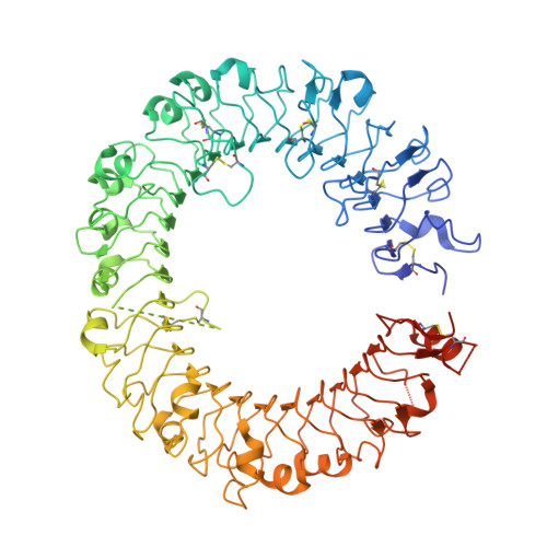
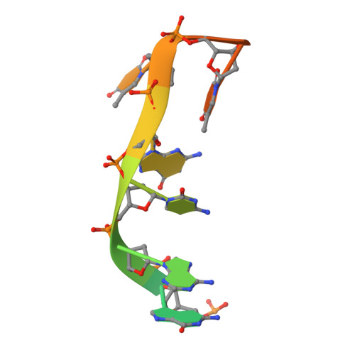
Structural basis of CpG and inhibitory DNA recognition by Toll-like receptor 9

Ohto, U.Shibata, T.Tanji, H.Ishida, H.Krayukhina, E.Uchiyama, S.Miyake, K.Shimizu, T.

(2015) Nature 520: 702-705

  • DOI: https://doi.org/10.1038/nature14138
  • Primary Citation Related Structures: 
    3WPB, 3WPC, 3WPD, 3WPE, 3WPF, 3WPG, 3WPH, 3WPI

  • PubMed Abstract: 

    Innate immunity serves as the first line of defence against invading pathogens such as bacteria and viruses. Toll-like receptors (TLRs) are examples of innate immune receptors, which sense specific molecular patterns from pathogens and activate immune responses. TLR9 recognizes bacterial and viral DNA containing the cytosine-phosphate-guanine (CpG) dideoxynucleotide motif. The molecular basis by which CpG-containing DNA (CpG-DNA) elicits immunostimulatory activity via TLR9 remains to be elucidated. Here we show the crystal structures of three forms of TLR9: unliganded, bound to agonistic CpG-DNA, and bound to inhibitory DNA (iDNA). Agonistic-CpG-DNA-bound TLR9 formed a symmetric TLR9-CpG-DNA complex with 2:2 stoichiometry, whereas iDNA-bound TLR9 was a monomer. CpG-DNA was recognized by both protomers in the dimer, in particular by the amino-terminal fragment (LRRNT-LRR10) from one protomer and the carboxy-terminal fragment (LRR20-LRR22) from the other. The iDNA, which formed a stem-loop structure suitable for binding by intramolecular base pairing, bound to the concave surface from LRR2-LRR10. This structure serves as an important basis for improving our understanding of the functional mechanisms of TLR9.


  • Organizational Affiliation
    • Graduate School of Pharmaceutical Sciences, The University of Tokyo, Hongo, Bunkyo-ku, Tokyo 113-0033, Japan.

Macromolecule Content 

  • Total Structure Weight: 92.94 kDa 
  • Atom Count: 5,783 
  • Modeled Residue Count: 720 
  • Deposited Residue Count: 813 
  • Unique protein chains: 1
  • Unique nucleic acid chains: 1

Macromolecules


Find similar proteins by:|  3D Structure
Entity ID: 1
MoleculeChains  Sequence LengthOrganismDetailsImage
Toll-like receptor 9801Bos taurusMutation(s): 0 
Gene Names: TLR9
UniProt
Find proteins for Q5I2M5 (Bos taurus)
Explore Q5I2M5 
Go to UniProtKB:  Q5I2M5
Entity Groups
Sequence Clusters30% Identity50% Identity70% Identity90% Identity95% Identity100% Identity
UniProt GroupQ5I2M5
Sequence Annotations
Expand
Reference Sequence
Find similar nucleic acids by:  Sequence
Entity ID: 2
MoleculeChains LengthOrganismImage
DNA (5'-D(*CP*AP*TP*GP*AP*CP*GP*TP*TP*CP*CP*T)-3')B [auth C]12synthetic construct
Sequence Annotations
Expand
Reference Sequence

Experimental Data & Validation

Experimental Data

  • Method: X-RAY DIFFRACTION
  • Resolution: 2.38 Å
  • R-Value Free:  0.274 (Depositor), 0.274 (DCC) 
  • R-Value Work:  0.217 (Depositor), 0.218 (DCC) 
  • R-Value Observed: 0.220 (Depositor) 
Space Group: C 1 2 1
Unit Cell:
Length ( Å )Angle ( ˚ )
a = 115.82α = 90
b = 113.08β = 100.61
c = 67.43γ = 90
Software Package:
Software NamePurpose
MOLREPphasing
REFMACrefinement
MOSFLMdata reduction
SCALAdata scaling

Structure Validation

View Full Validation Report


Entry History 

Deposition Data

Revision History  (Full details and data files)

  • Version 1.0: 2015-02-11
    Type: Initial release
  • Version 1.1: 2015-05-06
    Changes: Database references
  • Version 1.2: 2024-10-30
    Changes: Data collection, Database references, Structure summary