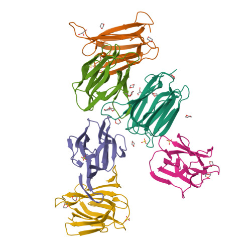
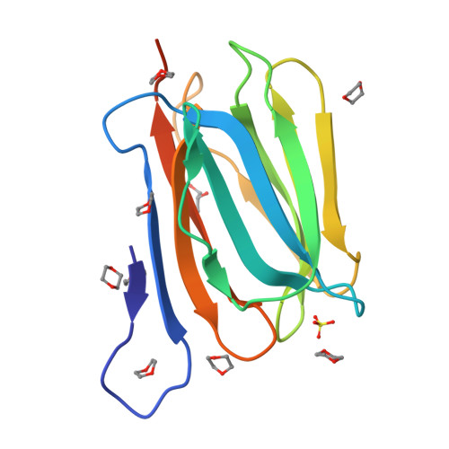
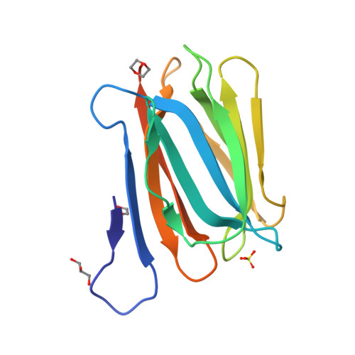
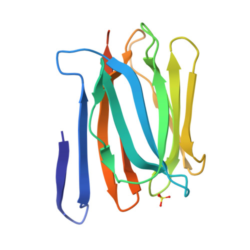
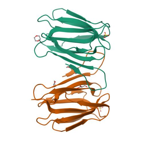
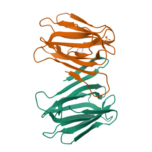
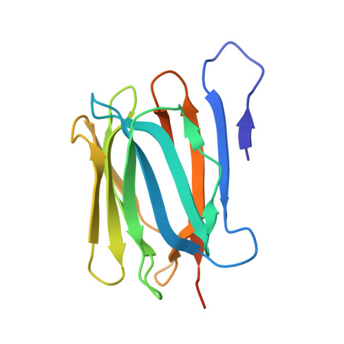
3WOC

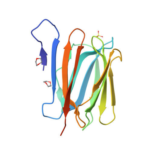
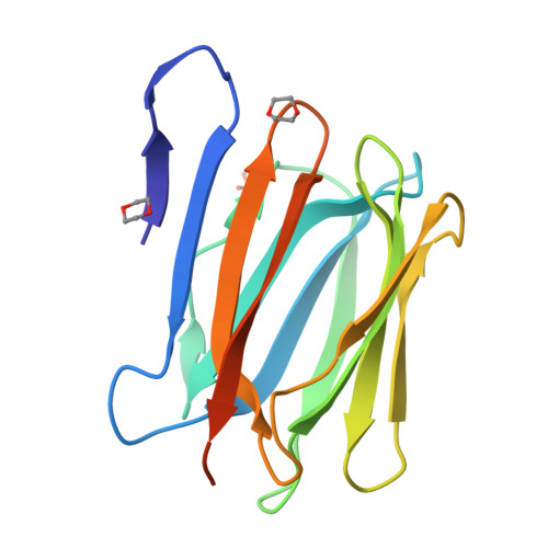
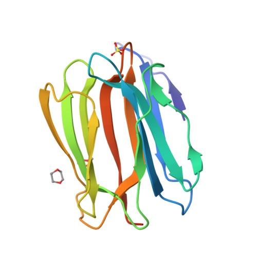
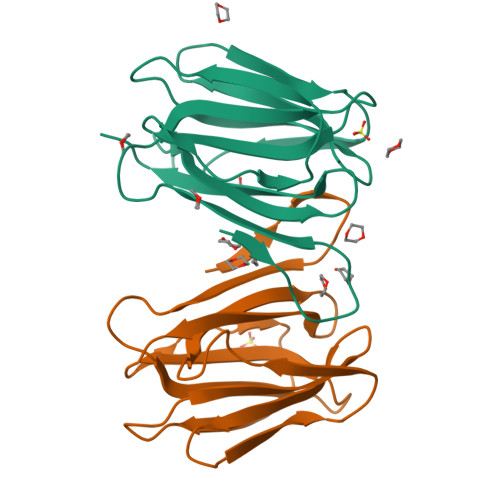
Crystal structure of a prostate-specific WGA16 glycoprotein lectin, form II


Experimental Data Snapshot

  • Method: X-RAY DIFFRACTION
  • Resolution: 2.40 Å
  • R-Value Free: 0.320 
  • R-Value Work: 0.252 
  • R-Value Observed: 0.256 

Starting Model: experimental
View more details

wwPDB Validation   3D Report Full Report


This is version 1.2 of the entry. See complete history


Literature

Discovery, Primary, and Crystal Structures and Capacitation-related Properties of a Prostate-derived Heparin-binding Protein WGA16 from Boar Sperm

Garenaux, E.Kanagawa, M.Tsuchiyama, T.Hori, K.Kanazawa, T.Goshima, A.Chiba, M.Yasue, H.Ikeda, A.Yamaguchi, Y.Sato, C.Kitajima, K.

(2015) J Biol Chem 290: 5484-5501

  • DOI: https://doi.org/10.1074/jbc.M114.635268
  • Primary Citation of Related Structures:  
    3WOB, 3WOC

  • PubMed Abstract: 

    Mammalian sperm acquire fertility through a functional maturation process called capacitation, where sperm membrane molecules are drastically remodeled. In this study, we found that a wheat germ agglutinin (WGA)-reactive protein on lipid rafts, named WGA16, is removed from the sperm surface on capacitation. WGA16 is a prostate-derived seminal plasma protein that has never been reported and is deposited on the sperm surface in the male reproductive tract. Based on protein and cDNA sequences for purified WGA16, it is a homologue of human zymogen granule protein 16 (ZG16) belonging to the Jacalin-related lectin (JRL) family in crystal and primary structures. A glycan array shows that WGA16 binds heparin through a basic patch containing Lys-53/Lys-73 residues but not the conventional lectin domain of the JRL family. WGA16 is glycosylated, contrary to other ZG16 members, and comparative mass spectrometry clearly shows its unique N-glycosylation profile among seminal plasma proteins. It has exposed GlcNAc and GalNAc residues without additional Gal residues. The GlcNAc/GalNAc residues can work as binding ligands for a sperm surface galactosyltransferase, which actually galactosylates WGA16 in situ in the presence of UDP-Gal. Interestingly, surface removal of WGA16 is experimentally induced by either UDP-Gal or heparin. In the crystal structure, N-glycosylated sites and a potential heparin-binding site face opposite sides. This geography of two functional sites suggest that WGA16 is deposited on the sperm surface through interaction between its N-glycans and the surface galactosyltransferase, whereas its heparin-binding domain may be involved in binding to sulfated glycosaminoglycans in the female tract, enabling removal of WGA16 from the sperm surface.


  • Organizational Affiliation

    From the Bioscience and Biotechnology Center, Nagoya University, Nagoya 464-8601, Japan.


Macromolecules
Find similar proteins by:  (by identity cutoff)  |  3D Structure
Entity ID: 1
MoleculeChains Sequence LengthOrganismDetailsImage
hypothetical protein
A, B, C, D, E
A, B, C, D, E, F
151Sus scrofaMutation(s): 0 
UniProt
Find proteins for A0A0A8IK66 (Sus scrofa)
Explore A0A0A8IK66 
Go to UniProtKB:  A0A0A8IK66
Entity Groups  
Sequence Clusters30% Identity50% Identity70% Identity90% Identity95% Identity100% Identity
UniProt GroupA0A0A8IK66
Sequence Annotations
Expand
  • Reference Sequence
Small Molecules
Ligands 3 Unique
IDChains Name / Formula / InChI Key2D Diagram3D Interactions
SO4
Query on SO4

Download Ideal Coordinates CCD File 
BA [auth F]
G [auth A]
Q [auth B]
U [auth C]
V [auth D]
BA [auth F],
G [auth A],
Q [auth B],
U [auth C],
V [auth D],
Y [auth E]
SULFATE ION
O4 S
QAOWNCQODCNURD-UHFFFAOYSA-L
GOL
Query on GOL

Download Ideal Coordinates CCD File 
P [auth A],
T [auth B]
GLYCEROL
C3 H8 O3
PEDCQBHIVMGVHV-UHFFFAOYSA-N
DIO
Query on DIO

Download Ideal Coordinates CCD File 
AA [auth E]
CA [auth F]
DA [auth F]
H [auth A]
I [auth A]
AA [auth E],
CA [auth F],
DA [auth F],
H [auth A],
I [auth A],
J [auth A],
K [auth A],
L [auth A],
M [auth A],
N [auth A],
O [auth A],
R [auth B],
S [auth B],
W [auth D],
X [auth D],
Z [auth E]
1,4-DIETHYLENE DIOXIDE
C4 H8 O2
RYHBNJHYFVUHQT-UHFFFAOYSA-N
Experimental Data & Validation

Experimental Data

  • Method: X-RAY DIFFRACTION
  • Resolution: 2.40 Å
  • R-Value Free: 0.320 
  • R-Value Work: 0.252 
  • R-Value Observed: 0.256 
  • Space Group: C 1 2 1
Unit Cell:
Length ( Å )Angle ( ˚ )
a = 134.483α = 90
b = 77.848β = 91.3
c = 87.187γ = 90
Software Package:
Software NamePurpose
HKL-2000data collection
MOLREPphasing
REFMACrefinement
DENZOdata reduction
SCALEPACKdata scaling

Structure Validation

View Full Validation Report


Entry History 

Deposition Data

Revision History  (Full details and data files)

  • Version 1.0: 2014-12-31
    Type: Initial release
  • Version 1.1: 2015-03-25
    Changes: Database references
  • Version 1.2: 2023-11-08
    Changes: Data collection, Database references, Derived calculations, Refinement description